Type
Characteristics
Injuries
Points
Skeletal/soft-tissue
Group
1
Low energy
Stab wounds, simple closed fractures, small-caliber gunshot wounds
1
2
Medium energy
Open or multiple-level fractures, dislocations, moderate crush injuries
2
3
High energy
Shotgun blast (close range) and high-velocity gunshot wounds
3
4
Massive crush
Logging, railroad, oil rig accidents
4
Shock
Group
1
Normotensive hemodynamics
BP stable in field and in OR
0
2
Transiently hypotensive
BP unstable in field but responsive to intravenous fluids
1
3
Prolonged hypotension
Systolic BP <90 mmHg in field and responsive to intravenous fluid only in OR
2
Ischemia
Group
1
None
A pulsatile limb without signs of ischemia
0a
2
Mild
Diminished pulses without signs of ischemia
1a
3
Moderate
No pulse by Doppler, sluggish capillary refill paresthesia, diminished motor activity
2a
4
Advanced
Pulseless, cool, paralyzed, and numb without capillary refill
3a
Ischemia
Group
1
<30 years
0
2
>30, <50 years
1
3
>50 years
2
In 1991, Russell et al. proposed the Limb Salvage Index (LSI) based on the review of 70 limb-threatening injuries. The index predicts the likelihood of limb salvage based on ischemia time and injury severity to six types of tissue that may be involved [63]. In order to specifically quantify each of these categories, extensive examination during an operation is necessary. The system is therefore very detailed and difficult to use in the acute decision-making process [5]. Another detailed scoring system, known as the NISSSA (nerve injury, ischemia, soft-tissue contamination, skeletal injury, shock, and age), was introduced by McNamara et al. in 1994. This system is a more complex modification of the MESS that separates the skeletal and soft-tissue injury, and adds a score for nerve injury. In a small retrospective series (24 patients), the authors concluded that the system is more sensitive and specific than the MESS [51].
13.5 Management
Initial management of the patient with a limb-threatening injury begins with ATLS protocol emphasizing a primary survey with immediate assessment of ABC’s (Table 13.2). Following this, the field dressing should be removed and any significant bleeding immediately controlled. This should be done with direct pressure, tourniquet, a compressive dressing, or proximal clamping (in that order of preference). Once the resuscitative effort is underway, further assessment of other injuries should be undertaken as well as a thorough neurovascular examination. If there is disruption to the arterial flow to the extremity, and salvage is being considered, an intraluminal shunt may be used. Wound dressing, gross alignment and splinting should be performed. Following this, any radiographic studies may be obtained (including vascular studies if necessary), and intravenous antibiotic and tetanus prophylaxis administered. We always calculate a MESS for each patient at the onset of treatment.
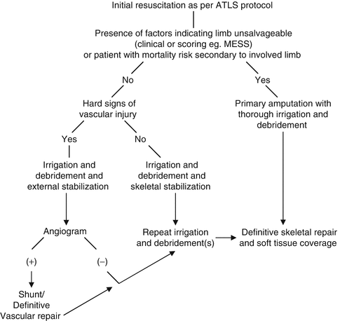
Table 13.2
Algorithm for the management of the patient with severe extremity trauma
If an early amputation is deemed necessary it is often advantageous to take medical record photographs to document the severity of the injury. Also, obtaining and documenting additional “attending” opinions on the need for the primary amputation is highly recommended. We also recommend keeping a photographic record throughout the course of treatment if reconstruction is performed, to document both progress and decline. Our indications for early amputation include unreconstructable osseous or soft-tissue injuries, irreparable vascular injuries, and severe loss of the plantar soft tissue (Fig. 13.1). Previous authors have recommended amputation if plantar sensation is absent. Recent evidence has suggested that initially absent plantar sensation does not predict a poor functional outcome, and that it may return in more than half of patients followed out to 24 months [8]. We therefore do not use absent plantar sensation as criteria for a primary amputation alone.
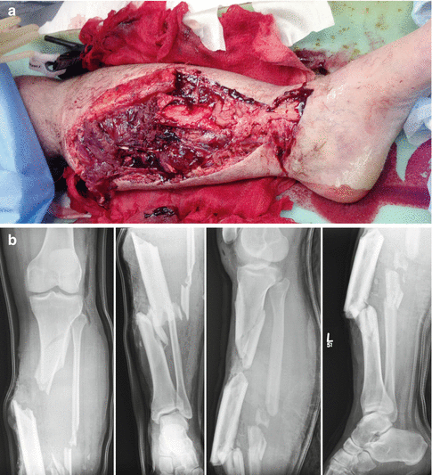
Fig. 13.1
A 25-year-old male sustained Gustillo and Anderson Type IIIB open tibia/fibula fractures following a crush injury in a motor cycle accident. (a) Clinical photo (top image) of the injury. (b) AP and lateral radiographs (bottom images) demonstrate a comminuted tibia and fibula fractures. The patient underwent early below-knee amputation secondary to unreconstructable soft tissue envelope
The amputation should be performed at the most distal level possible, but should not include clearly nonviable tissues. Examining color, consistency, contractility, and bleeding determine tissue viability. It has been shown that trans-tibial amputations have significantly better functional outcomes and lower energy expenditure than more proximal levels of amputation [18, 49]. A thorough irrigation and debridement should be performed without any attempt to close the wound at this time. A sterile dressing or wound VAC can be applied, and a splint placed if the amputation is below the level of the knee or elbow (Fig. 13.2). Return to the operating room with repeat surgical debridements should be performed as deemed necessary. In most instances several irrigation and debridements are undertaken prior to closure of the stump site to ensure adequate removal of nonviable tissue and a clean environment for wound healing.
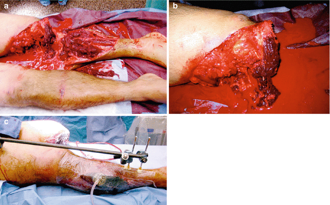
Fig. 13.2
A 21-year-old male presented to the emergency department following a motorcycle collision with bilateral lower extremity injuries. (a) Left-sided pulse-less (Grade IIIC) “mangled” knee/lower extremity injuries and a right-sided bicondylar closed tibial plateau fracture with compartment syndrome. (b) Left-sided completion of the above knee amputation retaining as much viable soft tissue as possible. (c) Application of negative pressure wound therapy dressing to left-sided amputation site, as well as external fixation of right bicondylar tibial plateau fracture and leg fasciotomies for compartment syndrome
If the need for amputation is not clear upon initial examination, then limb salvage should be attempted. Once again a thorough irrigation and debridement with removal of any contaminants and nonviable tissue performed emergently. This may be the most important operation the patient undergoes during the entire course of treatment. External fixation to gain stability of fractures and to aid in wound care is typically performed at this time. If necessary, a definitive vascular repair should be performed following skeletal stabilization. Ex-fix pins should be placed strategically away from the zone of injury and based on future incisions for definitive ORIF. Compromise of formal ORIF after DCO using external fixation is generally not an issue [68]. Fasciotomies should be performed as necessary. Antibiotic bead pouches and negative pressure wound therapy can be used to help decrease infection and assist with wound care [16, 37, 38, 54, 57]. The extremity is closely monitored over the next 24–72 h for soft tissue viability and sensorimotor function. Wounds should be regularly inspected, and repeat irrigation and debridements performed based on wound appearance (tissue viability, presence of contaminants, infection, etc.). VAC dressings are changed every 48–72 h.
If at any point the limb is deemed unsalvageable, or if the patient’s life is in jeopardy secondary to the extremity, amputation should be performed. If the extremity remains viable for reconstruction and the patient condition permits then definitive skeletal stabilization and early soft tissue coverage should be performed [28, 29]. The use of BMP-2 has been approved in complex open tibia fractures. It was shown to accelerate fracture healing, reduce infection rate, and decrease the need for secondary procedures to obtain union in a randomized, prospective study involving 450 open tibia fractures [32]. Further research involving a larger cohort of patients with longer follow-up is necessary to confirm these results, and analyze the long-term complications and outcomes. Some recent studies have shown increased rates of heterotopic ossification in peri-articular injuries [7], possibly increased risk of malignancies [13], and unreliable reporting of complications in the literature [14]. Until more data is available, the utility and safety of BMP in the setting of open fractures is still uncertain.
Various modalities are available for surgical fixation including uniplanar external fixators, hyrid external fixators, thin-wire ring external fixators, plate and screw constructs, and intramedullary nails. There are pros and cons of each modality. It is beyond the scope of this chapter to recommend the type of fixation to use in the setting of complex extremity trauma. Many patients will require further surgery to achieve osseous union and this should be discussed along with possible complications with each patient thoroughly [8, 29, 35] (Figs. 13.3 and 13.4).
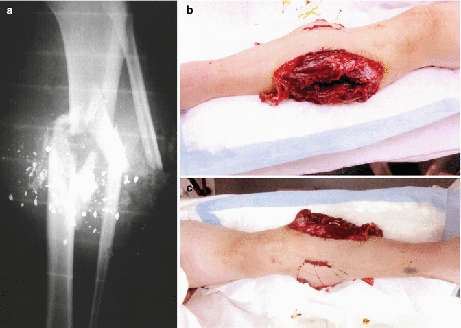
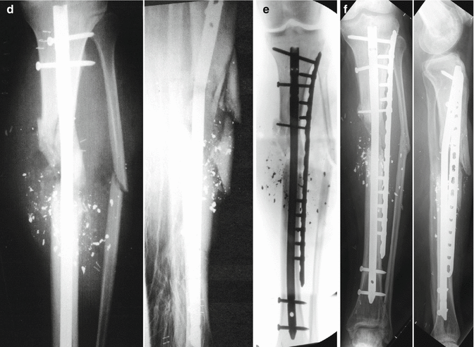
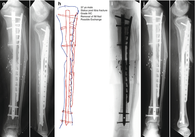
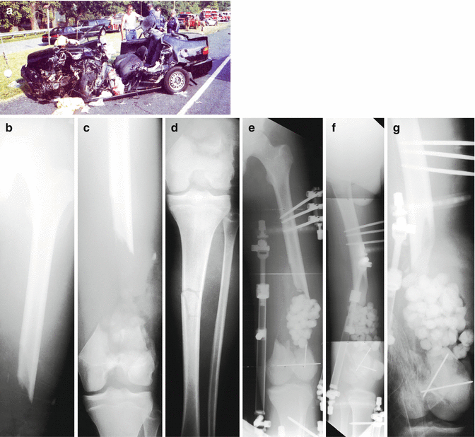
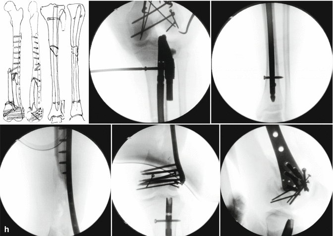
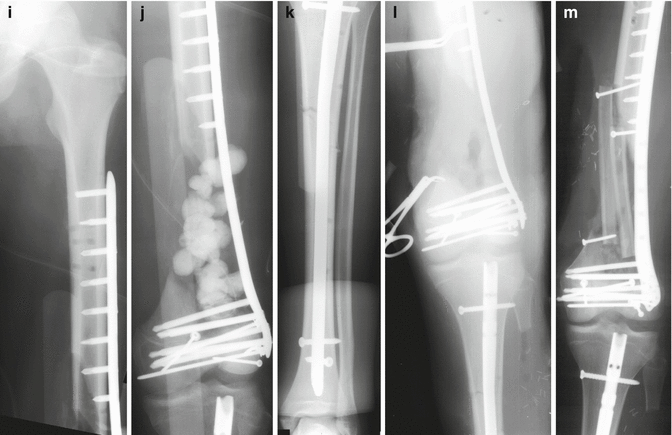
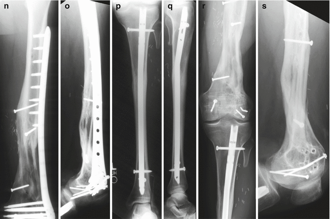
Fig. 13.3
A 36-year-old male was accidentally shot in the leg with a shotgun during a hunting trip. (a–c) He suffered an open, left-sided grade IIIC tibial shaft fracture with marked comminution. He also presented with complete functional deficit to his anterior compartment. He was taken to a local trauma center for irrigation and debridement (I&D), stabilization with and external fixation and a saphenous vein revascularization of the popliteal artery. Subsequent multiple I&D procedures were performed (including compromised bone). A negative pressure wound therapy dressing was placed over the wound sites. An inferior vena cava (IVC) filter was also inserted. (d) On day 3 a reamed, locked tibial intramedullary nail was inserted. (e) At 2 weeks following the injury, the patient was transferred to our institution for definitive management of his injuries. Repeat I&D was performed, the proximal interlocking screw was then removed to allow some correction of alignment and a percutaneous locking plate and screws was placed along the lateral surface of the tibia and a VAC dressing was applied. (g) Radiographs at 19 months illustrate some callus formation and a broken proximal interlocking screw. (h, i) Exchange IM nailing was planned and performed with placement of demineralized bone matrix (DBM) and a bone morphogenetic protein-7 (BMP-7) supplement. (j) At the latest follow-up visit at 29 months following revision surgery, he presented with good radiographic and clinical findings including increased callus formation and consolidation of the fracture, well-healed soft tissues, resolution of most pain symptoms, a return to activities of daily living and some recreational activities including weight training and skiing. A slight dorsiflexion lag was still present
Fig. 13.4
A 17-year-old male was involved in a head-on collision with a tractor trailer. After being trapped inside the vehicle for approximately 1 h, he was extricated and flown to a local trauma center. He was diagnosed with an open, Grade IIIC left-sided AO/OTA Type C3.3 distal femur fracture with segmental defect and an ipsilateral tibial shaft fracture. External fixation was placed for initial stabilization and antibiotic beads were subsequently placed in the defect at 3 days following injury. Open reduction and internal fixation (ORIF) was performed with placement of an intramedullary (IM) locked nail for treatment of the tibial shaft fracture and then ORIF of the distal femur fracture with placement of a less invasive stabilization system (LISS) locking plate and screws. One week later the antibiotic beads were removed and the defect was prepared for bone graft placement. A second incision was made along the lateral border of the ipsilateral fibula and a free vascularized fibula bone graft was harvested for transplant to the femoral defect. It was docked in a double barrel fashion and stabilized using screw fixation. Following surgery he returned for regular follow-up visits. Three months after surgery all of the fractures were healing with incorporation of bone graft. The LISS plate was removed 4.5 years following the initial surgery. The clinical and radiographic follow-up illustrated excellent results with bony union, full range of motion, and complete resolution of pain and return to pre-injury activities. (a) Photograph of the vehicle and the scene following the accident. (b–d) Anteroposterior (AP) X-rays illustrating an AO/OTA Type C3.3 distal femur fracture with segmental bone defect and an ipsilateral tibial shaft fracture. (e–g) AP and lateral radiographs following placement of external fixation and antiobiotic beads at the site of the segmental bone defect. (h) Counterclockwise from top-left; preoperative plan, fluoroscopic images showing placement of intramedullary nail for the tibial shaft fracture and locking screws and open reduction and internal fixation (ORIF) of the distal femur fracture with placement of a LISS locking plate and screws. (i–k) Immediate postoperative radiographs demonstrating adequate fixation and alignment (l) AP radiographs illustrating preparation of distal femoral bone defect for placement of vascular bone graft. (m) AP X-radiograph following free vascularized fibular bone and placement of screw fixation. (n–q) AP and lateral X-rays 3.5 years following ORIF showing healed a distal femur fracture with incorporation of the fibular bone graft and a healed tibial shaft fracture. (r, s) AP and lateral X-rays 8 months following removal of LISS plate and screws and 4.5 years following fracture surgery
13.6 Complications
A major factor in the decision making in the treatment of the mangled extremity is the possible major complications associated with the treatment arm chosen. Harris et al. reported the nature and incidence of major complications for patients enrolled in the LEAP study group. Their cohort consisted of 545 patients with severe lower-extremity injuries followed prospectively for 24 months. A physician examined each patient at 3-, 6-, 12-, 24-month intervals and major complications recorded. The two most common complications were wound infection (28.3 %) and nonunion (23.7 %), and the majority of each of these required operative intervention and inpatient care. Approximately a quarter of each of these complications were considered severe enough to compromise long-term function. The overall incidence of wound dehiscence was 8.6 % and that of osteomyelitis 7.7 %. There was also a 5.3 % incidence of symptomatic hardware [35]. The complication data from the cohort was further examined based on treatment arm in the study. A total of 149 patients underwent amputations, and the revision amputation rate was 5.4 %. The most common complications in this group were wound infection (34.2 %), followed by stump revision (14.5 %), phantom limb pain and wound breakdown (13.4 % each), and stump complications (10.7 %). In the limb reconstruction group the most common complication was nonunion (31.5 %), followed by wound infection (23.2 %). Of these infections 8.6 % developed into osteomyelitis. There was an incidence of post-traumatic arthrosis of 9.4 % and wound necrosis or breakdown of 6.5 %. The late amputation group (patients amputated after initial discharge) experienced the highest rate of major complications (85 %) [35].
This fact clearly highlights the need for appropriate decision making in the patient with a mangled extremity at the onset of treatment. Although there were no late mortalities reported, an incidence of up to 21 % has been reported in the literature. Bondurant et al. undertook an investigation looking at the effects of delayed versus primary amputation. There was a significant increase in length of hospital stay (22 versus 53 days) and number of surgical interventions (1.6 versus 6.9). The cost was almost double ($28,964 versus $53,462), and there was a 21 % mortality rate in the delayed amputation group [6]. It is quite evident that every effort should be made to avoid a late amputation given such high costs for all involved.
In a prospective cohort study (using LEAP study patients), Castillo et al. examined the specific effect of smoking on complication rate in severe open tibia fractures. A total of 268 patients with unilateral injuries were followed prospectively. Nonunion rates were significantly higher in both the current and previous smoking groups (37 % and 32 % respectively). The authors were able to demonstrate that current smokers were more than twice as likely to develop an infection, and 3.7 times more likely to have osteomyelitis. Previous smoking history was detrimental as well, and this group was 2.8 times more likely to develop osteomyelitis than nonsmokers. Their recommendation was that orthopedic surgeons should encourage patients to enter smoking cessation programs [15]. It has been shown that with the assistance of a surgeon up to 40 % of patients are able to quit smoking [58].
13.7 Predictive Ability of Scoring Systems to Predict Final Outcome
Some authors have examined the ability of the previously discussed scoring systems to predict functional outcome following treatment. Durham et al. performed a retrospective analysis of upper and lower severe extremity injuries to determine the validity and ability to predict outcome of the predictive indices discussed earlier. For each of the four systems analyzed, there were no significant differences between patients with good or poor functional outcomes [19]. Ly et al. reported on the ability of the five most commonly used predictive indices (above plus Hannover Fracture Scale-98) to determine functional recovery following limb salvage in a cohort of 507 patients (LEAP study group). The authors showed that none of the scoring systems analyzed were able to determine the outcome based on the SIP out to 24 months following injury [47]. One can conclude, based on these two studies, that the commonly applied predictive indices may be useful in early decision making, but are unable to predict functional recovery.
13.8 Outcomes Following Limb Salvage Versus Amputation
Recent medical and surgical technological advances have dramatically improved the surgeon’s ability to salvage severely injured extremities. Limbs that historically would have been amputated can now be managed with complex reconstruction techniques. Although the limb remains viable, it is often questioned whether or not the patient would have been better served with an amputation. Limb salvage patients often still complain of edema, loss of motion, pain, decreased sensation, difficultly with footwear and ambulation [41]. The end result is often a physical, psychological, financial, and social cripple with a useless salvaged limb [34, 36].
Hoogendorn and van der Werken looked at the long-term outcome and quality of life of patients treated with reconstruction versus amputation following Grade III open tibia fractures. A total of 64 patients were assessed, including 43 with successful limb salvage and 21 who underwent amputations (including both primary and delayed). Lower extremity impairment was determined using “Guides to the Evaluation of Permanent Impairment” of the American Medical Association. Quality of life was measured using the Nottingham Health Profile (NHP), the SF-36, and a questionnaire the authors specifically designed for the study examining pain, daily function, psychological factors, and handicap with working. Patients who underwent amputations had more severe injuries, and had a higher number of vascular injuries (77 % versus 17 %). The limb salvage group underwent more operations and had more complications [41].
Delayed amputations were performed in eight patients, most commonly secondary to persistent infection and poor soft tissues. They were hospitalized twice as long as those who underwent primary amputation. Others have shown that delayed amputation results in poorer functional outcome versus primary amputation [6, 25]. From the reported health surveys the authors found low scores in both groups but no significant differences. In both groups over half the patients considered themselves disabled, with a slightly higher percentage of patients who had amputations reporting difficulty with practicing a profession (60 % versus 40 %). Of particular interest was that the mean lower extremity impairment score was significantly worse for amputees (73.5 %) as compared to the limb salvage group (17.6 %). These patients therefore perceived a higher level of function than those who were amputated [41].
Related posts:


Stay updated, free articles. Join our Telegram channel

Full access? Get Clinical Tree


