Optical and Light-Guided Devices
John C. Sakles
Julie A. Slick
INTRODUCTION
Traditional laryngoscopes require the operator to achieve a direct line of site of the glottis by aligning the oral, pharyngeal, and laryngeal axes (Chapter 12). Video laryngoscopes capitalize on a video camera or chip, which is mounted on a specially designed blade, to circumvent the need for a straight line of sight, while providing superior views of the glottis and surrounding spaces (Chapter 13). Optically enhanced devices are those laryngoscopes that allow the operator to visualize the glottis without creating a straight line of sight, but without using video or fiberoptic technology. The glottic view is obtained by the use of inexpensive optics consisting of combinations of prisms, mirrors, and lenses. The relatively inexpensive cost of the technology is one of the major benefits of these devices when compared with the more expensive fiberoptic and video devices. The manufacturers of these devices also offer video capability through attachable video cameras, which are customized for use with individual optically enhanced laryngoscopes. The addition of video capability provides a magnified view of the glottis, and enhances education by allowing other providers to visualize what the airway manager is observing. There are only two true optical devices of relevance to emergency airway management, the Airtraq and the Truview.
DEVICES
Airtraq
Components
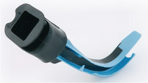
The Airtraq is a single-use, disposable laryngoscope that provides the operator with a magnified view of the glottic structures (Fig. 14-1). The Airtraq is now available in four sizes for orotracheal intubation. The orotracheal devices have an endotracheal tube (ETT) channel in which the ETT is preloaded before insertion. Airtraq also offers two devices that lack the ETT channel and are intended for use during nasotracheal intubation. These devices are intended for insertion through the mouth to facilitate nasotracheal intubations with the assistance of glottic visualization. They also offer a device with a larger channel that will accommodate double lumen endobronchial tubes. The Airtraq devices have a relatively narrow profile. The minimal mouth opening required is 18 mm for the the regular Airtraq (size 3) and 16 mm for the small Airtraq (size 2).
 Figure 14-1 • Airtraq Optical Laryngoscope. Note the black rubber eyepiece atop the viewing channel and blue endotracheal tube guide. Shown is the standard adult Airtraq, size 3. |
All of the Airtraq devices are made of plastic and are designed for single use. They cannot be cleaned or sterilized, and they will not function properly if this is attempted. This single use attribute makes the Airtraq particularly suitable for use in field settings for both emergency medical services (EMS) and military applications. The devices are powered by three AAA batteries, which will provide approximately 90 minutes of operating time. The shelf life is approximately 2 years. The Airtraq is turned on by pressing a button on the top of the unit that illuminates a low-heat lightemitting diode (LED). The device has a built-in antifog mechanism. A rubber eyepiece is connected to the optical channel. This rubber eyepiece can be removed and replaced with an optional video composite unit that will allow transmission of the optical image to a wired or wireless video monitor.
Operation
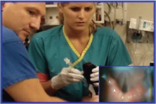
The Airtraq requires little setup time and is self-contained within its protective packaging. The Airtraq’s antifogging mechanism requires the device be turned on for approximately 30 seconds to maximize this benefit. The LED will flash when the device is turned on and become constant when the heating element has reached the appropriate antifog temperature and the device is therefore ready for use. The ETT should be lightly lubricated and preloaded into the ETT channel. No stylet is used. The anterior surface of the Airtraq blade, which will come in contact with the tongue, can be lubricated to help facilitate passing the device around the tongue. The Airtraq should be placed into the mouth using a midline approach. Gentle outward traction may be applied to the tongue using the operator’s free hand, if necessary, to ensure that the tongue is not pushed into the hypopharynx. As the Airtraq is advanced, the operator visualizes the epiglottis through the eyepiece and continues to move the device forward into the vallecula. When the vallecula is entered, the Airtraq is lifted in the vertical plane to elevate the epiglottis and align the vocal cords within the center of the optical field (Fig. 14-2). The ETT is slowly advanced through the vocal cords, and then disengaged from the device and the Airtraq is removed. If the ETT is obstructed by the epiglottis or arytenoids, the entire device is manipulated to better align the ETT position with the
glottic entrance. The Airtraq can be pulled back slightly and rotated along its longitudinal axis to help align the glottic structures. Alternatively, the epiglottis can be lifted directly by the Airtraq blade through a straight blade approach to appropriately align the glottic structures. However, this technique can somewhat distort the anatomy and is not the recommended approach.
glottic entrance. The Airtraq can be pulled back slightly and rotated along its longitudinal axis to help align the glottic structures. Alternatively, the epiglottis can be lifted directly by the Airtraq blade through a straight blade approach to appropriately align the glottic structures. However, this technique can somewhat distort the anatomy and is not the recommended approach.
If the proprietary composite video system is to be used, the rubber optical eyepiece is removed and the video adaptor is snapped onto the Airtraq before insertion. The operator then visualizes the entire procedure on the external monitor. This approach allows the operator to maintain a safe distance from the patient and provides a larger view of the anatomical structures.


