Monitoring and Documentation
Jeff Gadsden
Introduction
The incidence of complications from general anesthesia has diminished substantially in recent decades, largely due to advances in respiratory monitoring.1 The use of objective monitors such as pulse oximetry and capnography allows anesthesiologists to quickly identify changing physiologic parameters and intervene rapidly and appropriately.
In contrast, the practice of regional anesthesia has traditionally suffered from a lack of similar objective monitors that aid the practitioner in preventing injury. Practitioners of peripheral nerve blocks were made to rely on subjective end points to gauge the potential risk to the patient. This is changing, however, with the introduction and adoption of standardized methods by which to safely perform peripheral nerve blocks with the minimal possible risk to the patient. For example, instead of relying on feeling “clicks,” “pops”, and “scratches” to identify needle tip position, the anesthesiologist can now directly observe it using ultrasonography. It follows that advancements such as this may help in reducing the three most feared complications of peripheral nerve blockade: nerve injury, local anesthetic toxicity, and inadvertent damage to adjacent structures (“needle misadventure”).
Objective monitoring, and the rationale for its use, is discussed in the first part of this chapter. The later section focuses on documentation of nerve block procedures, which is a natural accompaniment to the use of these empirical monitors. The proper documentation of how a nerve block was performed has obvious medicolegal implications and aids the future practitioner in choosing the best nerve block regimen for that particular patient.
SECTION I: MONITORING
What Are the Available Monitors?
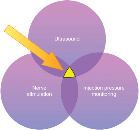
Monitors, as used in the medical sense, are devices that assess a specific physiologic state and warn the clinician of impending harm. The monitors discussed in this chapter include nerve stimulation, ultrasonography, and the monitoring of injection pressure. Each of these has its own distinct set of both advantages and limitations. For this reason, these three technologies are best used in a complementary fashion (Figure 5-1), to minimize the potential for patient injury, rather than just relying on the information provided by one monitor alone. The combination of all three monitors is likely to produce the safest possible environment in which to perform a peripheral nerve block.

FIGURE 5-1. Three modes of monitoring peripheral nerve blocks for patient injury. The overlapping area of all three (yellow area) represents the safest means of performing a block.
A fourth monitor that many clinicians use regularly is the use of epinephrine in the local anesthetic. Good evidence supports this practice as a means of improving safety during peripheral nerve blocks, particularly in patients receiving higher doses of local anesthetic. First, it acts as a marker of intravascular absorption. About 10 to 15 μg of epinephrine injected intravenously reliably increases the systolic blood pressure >15 mm Hg, even in sedated or beta-blocked individuals (whereas a heart rate increase is not reliable in sedated patients).2,3 The recognition of this increase permits the clinician to halt the injection promptly and increase his or her vigilance for signs of systemic toxicity. Second, epinephrine truncates the peak plasma level of local anesthetic, resulting in a lower risk for systemic toxicity.4,5 Concerns regarding the effects of epinephrine on nutritive vessel vasoconstriction and nerve ischemia have been unsubstantiated. In contrast, concentrations of 2.5 μg/mL have been associated with an increase in nerve blood flow, likely due to the predominance of the beta effect of the drug.6 Therefore, when added to local anesthetics, epinephrine can enhance safety during administration of larger doses of local anesthetics.
Nerve Stimulation
Neurostimulation has largely replaced paresthesia as the primary means of nerve localization in the 1980s and has only recently been challenged by ultrasound guidance. Its effectiveness as a method of nerve localization has been challenged since the publication of a series of studies showing that, despite intimate needle–nerve contact as witnessed by ultrasonography, a motor response may be absent.7 In some instances, a current intensity as high as >1.5 mA may be necessary to elicit motor response with needle placement within epineurium of the nerve.8 There are probably multiple factors that contribute to the explanation of this phenomenon, including the nonuniform distribution of motor and sensory fibers in the compound nerve and the unpredictable pattern of current dispersion in the tissue depending on tissue conductances and impedances.
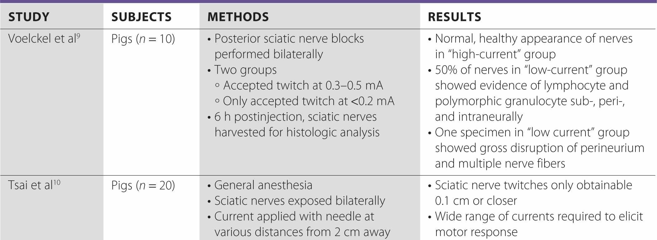
Although this has led some clinicians to de-value nerve stimulation in an era of ultrasound-guided blocks, a growing body of evidence suggests that the presence of a motor response at a very low current (i.e., <0.2 mA) is associated with an intraneural needle tip placement (Table 5-1). In a 2005 trial, Voelckel et al conducted percutaneous sciatic nerve blocks in pigs and demonstrated that when local anesthetic was injected at currents between 0.3 and 0.5 mA, the resulting nerve tissue showed no signs of an inflammatory process, whereas injections at <0.2 mA resulted in lymphocytic and granulocytic infiltration in 50% of the nerves.9 Tsai et al performed a similar study investigating the effect of distance to the nerve on current required; although a range of currents were recorded for a variety of distances, the only instances in which the motor response was obtained at <0.2 mA was when the needle tip was intraneural.10
TABLE 5-1 Studies of Intensity of the Current (mA) and Needle Tip Location


More recently, a study was conducted on 55 patients scheduled for upper limb surgery who received ultrasound-guided supraclavicular brachial plexus blocks. The authors set out to determine the minimum current threshold for motor response both inside and outside the first trunk encountered.11 They discovered that the median minimum stimulation threshold was 0.60 mA outside the nerve and 0.3 mA inside the nerve. Interestingly, stimulation currents of ≤0.2 mA were not observed outside the nerve, whereas 36% of patients experienced a twitch at currents <0.2 mA while the needle was intraneural.
Taken together, these data suggest that although the sensitivity of a “low-current” twitch for intraneural placement is not high, the specificity is. Put another way, the needle tip can be in the nerve and not elicit a motor response at very low currents; however, if a twitch is elicited at <0.2 mA, it is certain that the tip is intraneural.
Most regional anesthesiologists agree that injection of local anesthetic into the nerve may be a risk factor for injury and that extra-neural deposition minimizes the potential for an intrafascicular injection.12 Ultrasonography is good, but not perfect, at delineating the exact position of the needle tip. In our attempts to get “close, but not too close” to the nerve so we might have the best block result, needles occasionally but inevitably cross the epineurium into the substance of the nerve. This event in and of itself may be of minimal consequence.13 However, injection into a fascicle carries a high risk of injury.14 It is for this reason that a reliable electrical monitor of needle tip position is a useful safety instrument. If a motor twitch is elicited at currents <0.2 mA, our approach is to gently withdraw the needle until the motor response disappears and then attempt to reelicit the twitch at the more appropriate (0.3–0.5 mA) current.
Overall, nerve stimulation adds little to the cost of a nerve block procedure, in terms of time, clinician effort, or dollars. It also serves as a useful functional confirmation of the anatomic image shown on the ultrasound screen (e.g. “Is that the median or ulnar nerve?”). In our practice, nerve stimulator is routinely used in conjunction with ultrasound guidance as an invaluable monitor of the needle tip position with respect to the nerve, based on the association of low currents with intraneural placement. In addition, an unexpected motor response during ultrasound-guided blocks may alert the operator of the needle-nerve relationship that was missed on ultrasound.
Ultrasonography
The use of ultrasound guidance to assist in nerve block placement has become very popular, for a number of reasons. First, ultrasound allows visualization of the needle in real time and therefore quickly and accurately guide the needle toward the target. Multiple injection techniques that were difficult, or indeed dangerous, to do in the era of nerve stimulation alone are now easy to perform because the nerves can be seen and injectate carefully deposited at various points around them. Also, because a motor response is not technically required, blocks can now be performed in amputees who do not have a limb to twitch. Not surprisingly, ultrasound has the potential to improve the safety of peripheral nerve blocks for a number of reasons.
Stay updated, free articles. Join our Telegram channel

Full access? Get Clinical Tree


