Fig. 24.1
(a) Thoracic spine burst fracture of T6 with corpectomy and stabilization of body with titanium mesh gauge and small fragment plate. Spinal cord is exposed. (b) PLA membrane is fabricated to form posterior wall following corpectomy of T6. Mixture of autologous bone from vertebral body fracture and DBM putty is grafted in cage as well as anterior to the membrane which protects cord from bone graft spillage into spinal canal
The primary limiting factor in autologous bone transplant has been reported morbidity and complications associated with the harvest site as well as adequate volume for large defects. With defects of 2 cm or less, traditional anterior iliac crest bone graft is usually sufficient as 5–72 ml can be harvested [13]. Larger defects can still be grafted with iliac crest by multiple harvest sites such as the contra lateral site or use of the posterior iliac crests with amounts of 25–90 ml be obtained [13]. In addition, the use of a small acetabular reamer may result in less donor site pain and larger volume of graft [14].
The most recent development in autologous harvest techniques is the intramedullary canal harvest. A recent review confirms that the use of the Reamer Irrigator Aspirator (RIA) in a single pass reaming of the femur produces significant amounts of bone graft (25–90 ml) with low rates of complications and postoperative pain [13]. While the rate of complication is lower than that described in conventional iliac harvest, iatrogenic femur fracture has occurred. In addition, studies of RIA harvest material suggest that it is rich in growth factors, viable cells, and morselized trabecular bone [15]. The RIA harvest can thus be considered biologically equivalent to iliac graft. The bone marrow harvest, however, lacks any structural properties that can be achieved with tricortical iliac harvest.
In addition to autologous bone graft, bone graft substitutes can be utilized to augment the autograft harvest. Bone graft substitutes include osteoconductive materials such as synthetic tricalcium phosphates, calcium sulfates, and coral. These materials are fabricated as granules, blocks, strips, putties, and pastes. However, the efficacy of these materials as stand-alone graft in segmental defects is unknown [18]. Similarly, there are currently at least 40 commercial preparations of Demineralized Bone Matrix (DBM). DBM is an acid extract of human cadaveric bone consisting largely of type I collagen and other acid stable proteins including bone morphogenetic proteins. The osteoinductive content of the DBM is low and subject to the variables of donor biological activity, processing, and carrier [16]. The osteoconductive properties of the various commercial DBMs relate to carrier chemistry, adjunctive inorganic additives such as cadaveric cancellous bone or synthetic mineral. At present there are no prospective studies proving the benefits of DBM for the reconstruction of segmental bone defect. The primary use of DBM may be as an extender for autologous bone harvests such as intramedullary reaming harvest, cancellous bone, or marrow aspirates and concentrates [16]. The role of recombinant bone morphogenetic proteins in bone defect reconstruction continues to evolve [19]. The high cost, carrier characteristics, biological activity and mechanical qualities of available commercial BMP preparations limit its use at present mainly to small cortical defects and acute open tibia fractures.
There are numerous options for the application of the autologous bone graft. Defects up to 29 cm have been successfully grafted using the induced membrane technique as described recently by Masquelet [9]. At 4–6 weeks post-PMMA block implantation, the block is removed by longitudinally incising the encapsulating membrane. Autologous bone in the form of iliac graft or RIA bone marrow harvest, or autologous bone-bone substitute or autologous bone-allograft mixture is then used to fill the resulting cavity. A defect stabilized with an intramedullary nail will require less bone graft volume than defects stabilized with external fixation or plate constructs. A resorbable polylactide membrane can also be used to shape and contain the graft for applications such as the distal tibia and femur. In addition, resorbable membranes can be used to contain the graft in applications near the spinal cord, interosseous membrane of the forearm, or other applications where the reconstruction needs to be precisely configured [20]. The polymeric membrane may be used where bone grafting is done primarily such as the reconstruction of an unstable thoracic burst fracture where cancellous bone graft is combined with a titanium vertebral reconstructions cage and a posterior vertebral body wall is fabricated by molding a polymer membrane (Fig. 24.1). Another technique for applying autograft is the use of cylindrical titanium cages to form a weight bearing diaphysis. In this technique, titanium mesh cages that are typically used in spinal vertebral reconstructions are fashioned to bridge the defect which has been stabilized with an intramedullary nail. The cage is packed with cancellous bone and the cage–host bone margins are autografted to create a construct which has considerable immediate mechanical stability [21].
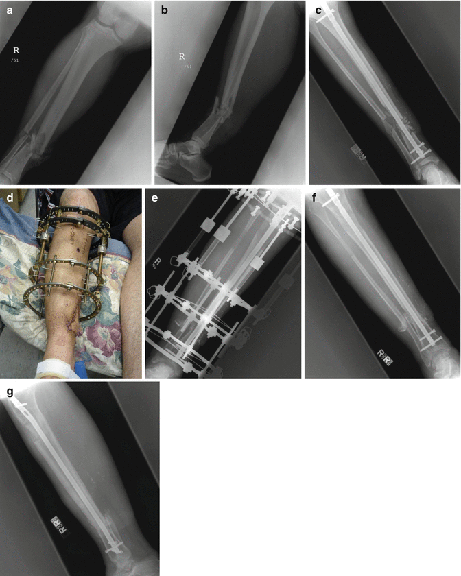
In summary, Phase III consists of bone reconstitution of the defect with an autologous bone graft. The autologous bone graft can consist of harvested iliac crest, intramedullary reaming harvests, or combinations of autogenous materials with synthetic bone substitutes or allograft materials. The stabilized defect can be prepared with the formation of an induced membrane, or bridged with a resorbable polylactide membrane or titanium mesh cage. Alternatively, Ilizarov distraction osteogenesis can be used, as shown in the case example (Fig. 24.2).


