Fig. 27.1
External fixator used as temporizing device awaiting soft tissue resolution in this multiply injured with severe midfoot fracture/dislocation. (a) Mid foot fracture/dislocation in multiply injured patient. (b, c) External fixator used as a temporizing device awaiting soft tissue resolution
27.4 Acute Total Orthopedic Care in the Poly-traumatized Patient with Associated Fracture
The breadth of research examining the poly-traumatized patient with associated long bone fractures has provided a foundation with which to further explore and refine treatment algorithms of orthopedic injuries in this patient population. The current approach to the poly-traumatized patient with skeletal fracture underscores the principle of early total care.
Early total care, when safe and feasible based on host and local conditions, affords the multiply-injured patient the best chances for long-term functionality. Long-term outcomes of poly-trauma survivors are often predicated on outcomes of associated musculoskeletal injury [13]. Thus, avoiding malunion and nonunion are of paramount importance for optimizing long-term outcomes after high energy trauma. Current care paradigm suggests that patients should receive definitive skeletal fixation once their systemic and local physiology can tolerate the required anesthesia, blood loss, and inflammatory response associated with operative fracture reduction and stabilization [14, 15].
27.5 Averting Malunion with an Optimized Initial Treatment Plan
The need for posttraumatic reconstruction is certainly inevitable in the poly-trauma patient population secondary to either nonunion, malunion or both. However, a primary goal of care in the acute setting is to prevent significant deformity leading to malunion. Early total fracture care in the poly-traumatized patient certainly minimizes the risk of developing malunion. Fracture reduction/instrumentation is drastically simplified and more reliable.
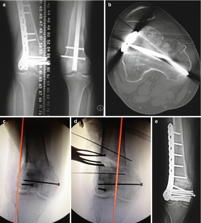
Underscoring the principle of averting malunion, symptomatic deformity correction is typically a complex posttraumatic reconstructive process (Fig. 27.2). Angular and/or rotational deformity requires corrective osteotomy. Operation through a contracted soft tissue envelope can lead to muscle imbalance or worse compromise of vital neurovascular anatomy. Further, even in the absence of major complication, outcomes after successful deformity correction can certainly be less than optimal in terms of ultimate patient outcome and satisfaction [16].


Fig. 27.2
A 20-year-old poly-trauma patient referred with significant malunion causing pain and ambulatory dysfunction. Malunion was associated with valgus and internal rotation deformity as well as shortening. Patella was chronically dislocated laterally. Complex posttraumatic reconstructive ladder was required, highlighting the consequences of marginal acute care. Osteotomy was performed to correct coronal, rotational. and length deformities. Further, soft tissue contracture release was necessary as well. Although the patient healed uneventfully, a 6-month course of healing/rehabilitation was required to achieve acceptable functionality. (a) Preoperative AP weightbearing radiograph. (b) Axial CT scan of right lower extremity. (c, d) Preoperative fluoroscopic image of the right knee with mechanical axis of the right lower extremity depicted in red. Intra-operative fluoroscopic image of the right knee with mechanical axis of the right lower extremity depicted in red. (e) Postoperative AP radiograph of right knee
27.6 Nonunion Reconstruction After Poly-trauma
When devising a treatment algorithm in the acute phase of injury, preventing skeletal fracture nonunion is of secondary importance relative to avoidance of deformity because of technical factors and physiologic impact of the procedures intended to correct these complications. From a technical standpoint, surgical corrective procedures for nonunion are less demanding for the treating orthopedic traumatologist and thus have a greater propensity to result in successful patient outcomes in contrast to analogous procedures for malunion.
For example, in the lower extremities, diaphyseal fractures of the tibia or femur that have been treated acutely with intra-medullary nailing but which have failed to unite can be treated with exchange nailing using a larger diameter rod [17]. If alignment and rotation of the long bone has already been restored during the index procedure, then this revision operation requires little technical consideration relative to an osteotomy procedure required for correction of malunion. Furthermore, reaming incites a healing response and provides local bone graft at the nonunion site while the minimal surgical exposure needed for surgical execution does not disrupt the local soft tissue vascularity.
For the small percentage of diaphyseal fractures that still do not heal with exchange intra-medullary nailing or similarly those periarticular fractures that have not united following the index formal reconstruction with various plating strategies, a revision operation consisting of a more extensive surgical dissection with plate application and utilization of biologic agents will likely be required. The objective under these circumstances is to provide additional mechanical stability as well as biologic factors to improve fracture healing potential.
In the case of nonunion after exchange nailing for a diaphyseal fracture, the plate spans the fracture site with the screws directed around the intra-medullary implant. Conversely, when nonunion is experienced at the site of a periarticular fracture, a supplemental plate must be applied within a new three-dimensional plane. In either situation, the surgical dissection is more extensive, however if the mechanical axis of the fracture is aligned and rotationally controlled then plate application and bone grafting imparts less stress to the local soft tissue environment and remains technically less demanding than correctional osteotomies formal-united fractures [18].
27.7 Biologic Supplementation for Nonunion After Poly-trauma
In addition to adding increased mechanical stability during nonunion reconstruction procedures, the biology within the local soft tissue environment must be enhanced in order to promote fracture healing in aseptic atrophic or oligotrophic cases. Most commonly non-structural cancellous autogenous or allogenic bone graft is incorporated into the fracture site to augment the local biology in these cases. Autogenous cancellous bone graft is osteogenic, osteoconductive, and osteoinductive and is therefore the gold standard grafting material. Bone graft from the iliac crest is preferred due to the large quantity of osseous tissue that can be extracted, and furthermore some researchers believe that iliac crest bone of intramembranous origin may be more osteoconductive than bone of enchondral origin [19]. Shortcomings of harvesting autologous bone graft include the limited amount of bone and complications at the donor site including infection and pain.
Recently, innovative methods have been devised to extract autlogous bone graft from the intra-medullary canal and condyles of long bones. This reamer irrigator aspirator (RIA) technique enables the extraction of significantly large volumes of autologous bone compared to iliac crest bone grafting therefore obviating the need to incorporate bone graft substitutes into nonunion surgery [20]. Additionally, bone graft from the femoral intra-medullary canal is believed to demonstrate similar osteogenic potential to autologous iliac crest bone graft. These devices have characteristics that are unique in comparison to traditional femoral reamers including cutting and suctioning designs that increase the risk for iatrogenic fracture and blood loss making it vital to fully comprehend these capabilities prior to use [21].
Allogenic bone graft functions primarily as an osteoconductive material that is typically used in conjunction with autologous bone graft materials to fill larger skeletal defects. Allogenic bone graft is advantageous because it is obtainable in greater quantity, however, it is less biologically active compared to autologous bone graft. Combining allograft cancellous bone with an osteoinductive agent is a common practice such as recombinanat bone morphogenic protein [22, 23]. However, the relative osteoinductive capability must be weighed against the potential harmful side effects including carcinogenic risk [24].
27.8 Strategic Bone Grafting Approaches
Achieving bone graft incorporation and uneventful nonunion resolution is both art and science. The desirable mechanical environment typically includes rigid nonunion stabilization to allow for bone graft healing via creeping substitution. The soft tissue milieu necessary includes a well vascularized muscular sleeve.
The nonunion site should be debrided to healthy margins removing all interposed scar and necrotic bone ends. Stimulation of the nonunion site is effective by using the feathering method with either osteotome, drill or low speed bur. If fracture geometry is conducive without excessive shortening, bone graft is applied at the nonunion site and mechanical compression is maintained.
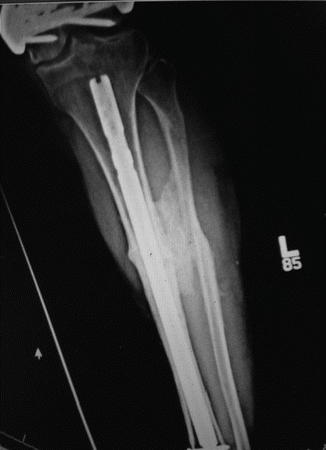
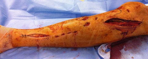
Besides direct application of bone graft to the nonunion, autograft and/or allograft/bmp can be used more creatively. For instance, tibia nonunion in the setting of an intact fibula is a desirable indication for creation of a surgical synostosis in an effort to restore long-term skeletal stability via creation of a one-bone leg (Fig. 27.3). Posterolateral grafting is ideally suited for diaphyseal tibial nonunion whereby the cancellous graft is applied along the well vascularized interosseous membrane below the posterior compartment musculature. For distal third fractures, a central bone grafting technique has been popularized [25].


Fig. 27.3
Previous poly-trauma patient with Grade 3b open tibia fracture with subsequent nonunion refractory to exchange nailing. Posterolateral bone grafting strategy performed creating a so-called one bone leg
In contrast, creation of a surgical synostosis would not be an effective strategy for radius and/or ulna nonunion. Functionality secondary to restricted forearm rotation would make this strategy less than ideal. Therefore, bone graft containment can be achieved with placement of a rigidly applied tricortical graft.
27.9 Open Fracture with Bone Loss: “Expected Nonunions”
The acute total care principles can be further applied to the poly-traumatized patient sustaining high grade open fracture with bone loss. Aggressive open fracture care is indicated to remove septic nidus yet typically bone defect is a result. The initial aim of the osseous reconstruction is to restore the alignment so as to prevent development of deformity. For diaphyseal fractures of the lower extremities, intra-medullary nailing is the standard care paradigm for rapidly establishing rigid internal splinting. In contrast, periarticular fractures are typically stabilized with fixed angle plate osteosynthesis (Fig. 27.4). Intra-articular fractures are precisely aligned then bridged to the shaft with appropriate length, alignment, and rotation [26]. Rigid stability of the fracture provided by the internal fixation will not only stop the cycle of ongoing injury but also allow for early patient comfort, mobility, and early physiotherapy during the subacute recovery phase from the trauma.


Fig. 27.4
Minimally invasive bridge plating of comminuted periarticular fracture restores alignment preventing deformity while preserving fracture biology and encouraging bony union
“Expected nonunions” after severe open fractures with bone loss are more easily managed in the absence of concomitant deformity. A proven methodology is usage of the Masquelet technique by initially filling large bone defects with antibiotic beads to encourage wound sterilization and neovascularization [27]. The antibiotic beads or spacer fills the void left by the removal of devitalized bone and prevents scar formation at the site of bone defect. Antibiotics are eluded from the porous cement architecture to aid in preventing infection within the local bone/soft tissue environment. Further, the beads incite an inflammatory process creating a vascular pocket ideally suited for future bone graft incorporation [28].
Stay updated, free articles. Join our Telegram channel

Full access? Get Clinical Tree


