Lumbar and sacral plexus anatomy

CHAPTER 23 Lumbar and sacral plexus anatomy


The anterior divisions of the lumbar, sacral, and coccygeal nerves form the lumbosacral plexus. The plexus is usually divided into three parts: the lumbar, sacral, and pudendal plexuses, for ease of description. The lumbar plexus primarily innervates the ventral aspect, whereas the sacral plexus innervates the dorsal aspect of the lower limb.


Lumbar plexus


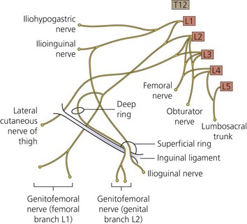
The lumbar plexus (Fig. 23.1) lies deep within the psoas major muscle in front of the transverse processes of the lumbar vertebrae. It is formed by the ventral rami of the first three lumbar nerves and the greater part of the ventral ramus of the fourth nerve. All the branches of the plexus emerge from the substance of the psoas major.


The first lumbar nerve, frequently supplemented by the 12th thoracic, splits into an upper and a lower branch; the upper and larger branch divides into the iliohypogastric and ilioinguinal nerves, the lower and smaller branch unites with a branch of the second lumbar to form the genitofemoral nerve.


The remainder of the second nerve, and the third and fourth nerves, divide into ventral and dorsal divisions. The ventral division of the second unites with the ventral divisions of the third and fourth nerves to form the obturator nerve. The dorsal divisions of the second and third nerves divide into two branches, a smaller branch from each uniting to form the lateral cutaneous nerve of the thigh, and a larger branch from each joining with the dorsal division of the fourth nerve to form the femoral nerve.


The femoral nerve


The femoral nerve, the largest branch of the lumbar plexus, arises from the dorsal divisions of the second, third, and fourth lumbar nerves. It descends through the fibers of the psoas major, emerging from the muscle at the lower part of its lateral border, and passes down between it and the iliacus muscle, behind the iliac fascia; it then runs beneath the inguinal ligament into the thigh, and splits into an anterior and a posterior division. In the thigh, the anterior division of the femoral nerve gives off anterior cutaneous and muscular branches. The anterior cutaneous branches comprise the intermediate and medial cutaneous nerves.


The intermediate cutaneous nerve pierces the fascia lata (and generally the sartorius) about 7.5 cm below the inguinal ligament, and divides into two branches that descend in immediate proximity along the forepart of the thigh to supply the skin as low as the front of the knee.


The medial cutaneous nerve passes obliquely across the upper part of the sheath of the femoral artery, and divides in front or at the medial side of that vessel into two branches: an anterior and a posterior. The anterior branch runs downward on the sartorius, perforates the fascia lata at the lower third of the thigh, and divides into two branches. The posterior branch descends along the medial border of the sartorius muscle to the knee, where it pierces the fascia lata, communicates with the saphenous nerve, and gives off several cutaneous branches.


The posterior division of the femoral nerve gives off the saphenous nerve, and muscular and articular branches.


< div class='tao-gold-member'>

Stay updated, free articles. Join our Telegram channel

Jul 28, 2016 | Posted by in ANESTHESIA | Comments Off on Lumbar and sacral plexus anatomy

Full access? Get Clinical Tree

Get Clinical Tree app for offline access