CHAPTER 28 Iliacus block
Clinical anatomy
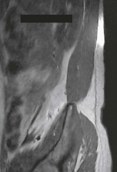
The iliacus fascia covers the iliacus and psoas muscles in the pelvis and descends into the thigh with these muscles (Fig. 28.1). The femoral nerve lies anterior to the psoas muscle initially, with the lateral cutaneous nerve of the thigh lateral to the psoas muscle and obturator nerve medial. At the inguinal ligament, the femoral nerve lies in a gutter between the psoas and iliacus muscles. These nerves thus lie beneath the iliacus fascia (Fig. 28.1). Spread of local anesthetic (Figs 28.2 and 28.3) beneath the iliacus fascia produces a higher success rate of anesthesia of the femoral nerve, lateral cutaneous nerve of the thigh, and obturator nerves than the femoral nerve block technique.
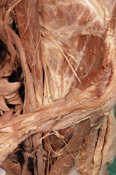
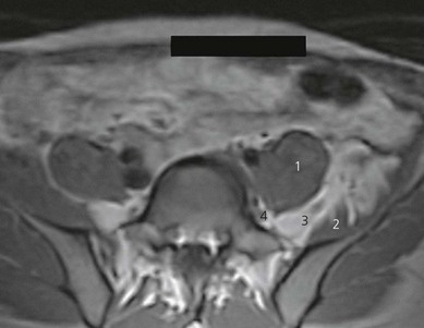
Figure 28.2 Axial T1-weighted MR image after injection of 40 mL of contrast, showing spread of injectate. Compare with Figure 21.5. Note contrast surrounding femoral and obturator nerves. Spread is via the plane between the iliacus and psoas muscles. 1: psoas muscle; 2: iliacus muscle; 3: femoral nerve; 4: obturator nerve.
Surface anatomy
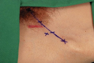
The inguinal ligament is outlined by a line connecting the anterior superior iliac spine and the pubic tubercle. The inguinal ligament is divided into equal thirds. At the junction between the outer one-third and inner two-thirds, a perpendicular line is drawn; 1 cm along this line is the needle insertion point (Fig. 28.4). The femoral artery can be palpated 2–3 cm more medially in the groin.
< div class='tao-gold-member'>
Related posts:


Stay updated, free articles. Join our Telegram channel

Full access? Get Clinical Tree


