Fig. 23.1
(a) Anatomy of the shoulder joint. (1) Humerus, (2) scapula, (3) articular capsule, (4) tendon of the biceps brachii muscle, (5) subscapular muscle, (6) acromion, (7) coracoid process (With permission from Danilo Jankovic). (b) Shoulder joint. Articular cavity and articular capsule. (1) Articular capsule, (2) articular cavity, (3) scapula, (4) acromion (With permission from Danilo Jankovic)
Strictly aseptic conditions are necessary when carrying out intra-articular injections.
Materials
25-G needle 30–40 mm long, 2- and 5-mL syringes, sterile swabs, disinfectant, sterile gloves, and sterile drape.
Ventral Access Route
Technique
The patient is seated with the supinated arm hanging freely, and the articular cavity is palpated directly medial to the head of the humerus. The needle is introduced underneath the clavicle, directly lateral to the coracoid process toward the outside and back. The path to the joint is very short with this approach (Fig. 23.2).
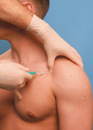
Fig. 23.2
Intra-articular injection into the shoulder joint from the ventral access route (With permission from Danilo Jankovic)
Dosage
2 mL local anesthetic—e.g., 0.5–0.75 % ropivacaine or 0.25 % bupivacaine mixed with 40 mg methylprednisolone.
Dorsal Access Route
Landmarks (Fig. 23.1a, b)
Spine of the scapulaRelated posts:

Stay updated, free articles. Join our Telegram channel

Full access? Get Clinical Tree


