Fiberoptic and Video Intubating Stylets
Valerie A. Dobiesz
Calvin A. Brown III
John C. Sakles
INTRODUCTION
Fiberoptic and video intubating stylets are novel intubating devices that permit visualization of the glottis by way of an image conveyed to an eyepiece or video screen from a distally positioned video or fiberoptic image source. Therefore, they do not require a direct line of sight from the operator’s eye to the glottis, as must occur for successful direct laryngoscopy. Distinct from the video laryngoscopes (see Chapter 13) and optical devices (Chapter 14), the fiberoptic and video stylets are intended to have the endotracheal tube (ETT) mounted directly over them, as for any conventional stylet, and are used to guide the tube through the cords and into the trachea under continuous visualization. Unlike flexible fiberoptic devices, these devices are rigid or semirigid and include a fiberoptic bundle or video apparatus enclosed in a preformed, curved steel stylet designed to navigate around the tongue and traverse the hypopharynx to visualize laryngeal structures, often with minimal mouth opening or neck mobility. This carries significant advantages; anatomical impediments to direct laryngoscopy, such as a high larynx, cervical spine immobility, or limited mouth opening often create little or no difficulty. Also, rigid stylets do not have any control mechanisms like their flexible fiberoptic counterparts and thus typically are easier to maneuver, especially for nonexperts. Rigid stylets have nonmalleable curved metal sheaths, the shape of which cannot be altered, whereas semirigid devices, although not flexible, can be bent slightly to alter their angulation and thus fit the particular airway geometry of each patient.
Semirigid fiberoptic stylets include the Shikani optical stylet (SOS) and the Levitan/“first pass success” (FPS) scope (Clarus Medical, Minneapolis, MN). The predominant semirigid video stylet is the Clarus Video System (CVS). Rigid stylets include the Bonfils Retromolar Intubation Fiberscope (Karl Storz Endoscopy, Tuttlingen, Germany) and the Video Rigid Flexible Laryngoscope (RIFL) (AI Medical Devices, Inc., Williamston, MI). New intubating stylets, all similar in shape and principle, are appearing on the market at regular intervals.
Although these devices are not yet a routine part of emergency airway management, they have shown significant potential as adjunctive devices for the difficult airway especially when mouth opening is limited, as a method of awake intubation, or as rescue devices for the failed airway. Their clear advantages over direct laryngoscopy suggest that they will come into increasing use as an airway management tool, even for “routine” emergency airways. They also may serve an expanding role in airway training because they all have video displays or are easily adapted for video by attachment of an eyepiece video camera adapter that transmits images to a video monitor.
The prototypical fiberoptic intubating stylet is the SOS; therefore, more time is devoted to its description, proper use, advantages, and contraindications. The other devices are similar in their core design and application, and are therefore described in less detail, highlighting specific features and differences.
SEMIRIGID STYLETS
Shikani Optical Stylet
The SOS is a semirigid stylet containing fiberoptic bundles for light and image transmission (Fig. 16-1). The stylet, rounded distally to an angle of about 70° to 80°, ends proximally in a highresolution, fixed-focus eyepiece. The adult stylet can accommodate ETTs of 5.5-mm internal diameter (ID) or larger. A pediatric version is available and accommodates tubes of 3.0- to 5.0-mm ID. A bright halogen light is powered from the attached handle, which holds four AA batteries, but the stylet is also compatible with green-specification fiberoptic laryngoscope handles or remote light sources through a fiberoptic cable. A camera can be attached to the eyepiece and the image displayed on a video monitor for teaching purposes. A push-button power switch can be found on the top of the handle. An adjustable tube stop is mounted on the proximal portion of the stylet to hold the ETT in the desired position and prevents the tip of the stylet from protruding from the distal end of the ETT. The tube stop incorporates an oxygen port, permitting insufflation of
oxygen. This helps prevent contamination of the tip of the stylet and can mitigate oxygen desaturation during prolonged intubation attempts. The malleable distal section of the stylet can be adjusted by hand, increasing or decreasing the angle of the bend to conform to the patient’s anatomy. The manufacturer also sells a stylet-bending guide that helps to bend the stylet in a smooth curve and helps avoid damage to the fiberoptic bundles by bending it too acutely.
oxygen. This helps prevent contamination of the tip of the stylet and can mitigate oxygen desaturation during prolonged intubation attempts. The malleable distal section of the stylet can be adjusted by hand, increasing or decreasing the angle of the bend to conform to the patient’s anatomy. The manufacturer also sells a stylet-bending guide that helps to bend the stylet in a smooth curve and helps avoid damage to the fiberoptic bundles by bending it too acutely.
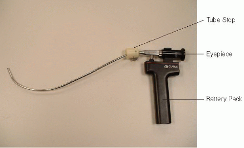 Figure 16-1 • Shikani Optical Stylet. |
To prepare the SOS, an ETT is loaded on the stylet, with the distal end of the stylet positioned just proximal to the ETT tip, and stabilized in this position by adjusting the tube stop proximally. Lubrication of the stylet will facilitate tube withdrawal when the intubation is completed, but care should be taken not to contaminate the tip of the scope, which will obscure the image. Before insertion, the stylet tip should be warmed with either warmed saline or a warm blanket. It is highly recommended that antifog solution be applied to minimize fogging during the intubation attempt. The device is held by the fingertips and thumb of the dominant hand, with the handle cradled in the web space between the thumb and index finger and the pads of all other fingers resting on the anterior part of the eyepiece and proximal stylet (Fig. 16-2). Despite its appearance, the handle should not be gripped in the hand, but is properly held in the fashion demonstrated in Figure 16-2. The ETT-stylet combination is then inserted into the mouth in the midline and advanced into the hypopharynx under direct vision, not by using the eyepiece. The entire stylet is oriented in the midline and advanced along its curve gently around the base of the tongue. A firm jaw lift/tongue pull during insertion will lift the soft tissues of the upper airway and create some anatomical space through which to navigate the scope. The operator should begin to visualize glottic structures through the eyepiece as the tip navigates the base of the tongue. The epiglottis should quickly come into view. Guide the stylet under the epiglottis to visualize the laryngeal inlet. The operator should then attempt to advance the tip of the stylet through the cords. Typically the operator can advance the scope 1 to 2 cm into the laryngeal inlet. A common error is to advance the fiberoptic tip too far into the hypopharynx as it is inserted, giving a view of the posterior aspect of the hypopharynx or upper esophagus. To avoid this, ensure that the primary motion of the scope is initially rotation around the tongue and not advancement into the hypopharynx. If no anatomical structures are recognized on the initial insertion, it is best to withdraw the stylet, ensure positioning in the midline, ensure proper elevation of the tongue and mandible, and attempt slow reinsertion, identifying the epiglottis or other laryngeal structures as the assembly is advanced. As with other intubating stylets, the instrument can be used in conjunction with direct laryngoscopy. For example, when an unanticipated grade 3 direct laryngoscopy (epiglottis only) occurs, the ETT-SOS stylet combination can be inserted under the epiglottis during direct laryngoscopy, and the glottic opening can then be located by looking through the eyepiece. With either technique, the
ETT-stylet is advanced through the cords as the operator looks through the eyepiece, and then the ETT is held in place as the stylet is withdrawn by the operator, using a large circular motion, in an arc initially upward toward the ceiling in the axis of the proximal end of the tube, and then continuing toward the patient’s chest and feet, following the curve of the stylet to facilitate removal (Fig. 16-3). Tube placement confirmation is with end-tidal carbon dioxide (CO2), auscultation, and chest radiography, as for any other methods of intubation.
ETT-stylet is advanced through the cords as the operator looks through the eyepiece, and then the ETT is held in place as the stylet is withdrawn by the operator, using a large circular motion, in an arc initially upward toward the ceiling in the axis of the proximal end of the tube, and then continuing toward the patient’s chest and feet, following the curve of the stylet to facilitate removal (Fig. 16-3). Tube placement confirmation is with end-tidal carbon dioxide (CO2), auscultation, and chest radiography, as for any other methods of intubation.
The SOS is advertised as being useful for the management of difficult and routine airways, with the video capability facilitating airway management teaching. In the teaching setting, coupling the device with a video system can greatly enhance success by allowing the instructor to help guide and, if necessary, reorientate the learner.
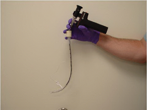 Figure 16-2 • Shikani Optical Stylet—in Hand. |
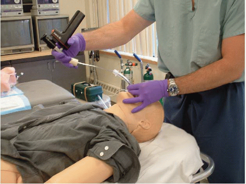 Figure 16-3 • Removal of a Shikani Optical Stylet. |
The primary limitation of the SOS is its inability to maintain clear vision occasionally because of fogging or the presence of secretions or blood. Fogging can be greatly reduced by warming the lens and applying antifog solution, as described previously. Although secretions, vomitus, or blood can obscure the distal lens of the scope, two key design elements come into play:
1. The patient is typically supine, and with the jaw thrust and tongue pull, most of the manipulation of the scope is occurring anterior to the location of the pooled liquids.
2. If the lens becomes obscured and cannot be cleared, it is quickly and easily removed, wiped, and reinserted in a matter of seconds.
Occasionally, the glottis cannot be visualized using the scope, and in such cases, adjustment of the scope’s curvature (usually increasing the angle, but sometimes decreasing it) provides an improved “angle of attack.”
Clarus Video System
The Clarus Video System (Clarus Medical, Minneapolis, MN) is the latest incarnation of the SOS (Fig. 16-4). It is the same shape and design as the SOS but has some new features that make it a superior device. Most importantly, a camera and a 4 in (10 cm) liquid crystal display (LCD) screen replace fiberoptic bundles and the eyepiece on the SOS. The stylet is more malleable at the end than the SOS, making it adjustable for individual airway anatomy. The stylet can be removed from the handle and display unit making it efficient to clean and disinfect. During intubation, the operator stands upright and looks at the screen instead of having to bend forward with the eye opposed to the eyepiece. The screen swivels, making it easy for the operator to adjust the viewing angle during the intubation. Another novel feature of the Clarus VS is the presence of a red light-emitting diode (LED) on the tip of the fiberoptic stylet. This provides transillumination through the soft tissues of the anterior neck during intubation and thus can potentially be useful as a guide or “locator” if the lens gets contaminated and the operator cannot see the airway. The Clarus VS in effect can be used like a lighted stylet, with tracheal entry being signified by a strong glow of red light through the anterior neck. The Clarus VS also has a video out port allowing the operator to display the image of the airway on a larger monitor or record the intubation using a video recorder. Instead of using disposable batteries like the SOS, the Clarus VS uses an internal rechargeable battery system that provides hours of power under typical conditions.
Levitan/FPS Scope
The Levitan/FPS scope is a small, semirigid fiberoptic stylet intended to be used in concert with a standard laryngoscope. Its preformed shape is similar to other intubating stylets but with a gentler
curve (approximately 45°); however, it can be gently bent to meet the unique geometry of most airways (Fig. 16-5). It has a handle with a small battery pack that powers an LED to provide illumination. A screw-down cap, rather than a push button, is used to activate the light source. The eyepiece is at the proximal end of the stylet, near the top of the battery pack. The mechanics of the Levitan/FPS are similar to that of the SOS with one important difference: the Levitan/FPS is intended


curve (approximately 45°); however, it can be gently bent to meet the unique geometry of most airways (Fig. 16-5). It has a handle with a small battery pack that powers an LED to provide illumination. A screw-down cap, rather than a push button, is used to activate the light source. The eyepiece is at the proximal end of the stylet, near the top of the battery pack. The mechanics of the Levitan/FPS are similar to that of the SOS with one important difference: the Levitan/FPS is intended
Related posts:


Stay updated, free articles. Join our Telegram channel
Full access? Get Clinical Tree
