, Corinna Eleni Psomadakis2 and Bobby Buka3
(1)
Department of Family Medicine, Mount Sinai School of Medicine Attending Mount Sinai Doctors/Beth Israel Medical Group-Williamsburg, Brooklyn, NY, USA
(2)
School of Medicine Imperial College London, London, UK
(3)
Department of Dermatology, Mount Sinai School of Medicine, New York, NY, USA
Keywords
Digital mucous cystGanglion cystMucoidMyxoidPseudocystDistal interphalangeal jointProximal nail foldNail changeTransilluminateExcisionIncision and drainageI&DCryosurgeryElectrodesiccation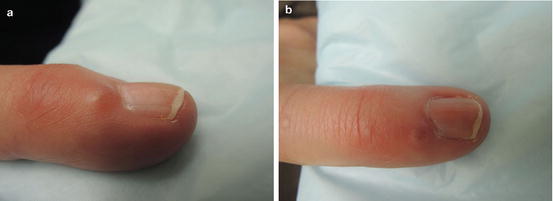
Fig. 8.1
(a and b) Proximal nail fold with firm, smooth subcutaneous nodule
Primary Care Visit Report
A 59-year-old female with no past medical history presented with a lesion on the pinky finger of her right hand. She first noticed the sore about 2–3 months prior. It was not painful, but it had started to distort her fingernail.
Vitals were normal. On exam, on the right hand fifth digit, just proximal to the nail bed, there was a 2 mm soft, non-tender, fluctuant mass with nail deformation.
This was treated as a digital mucous cyst , and the patient was referred to dermatology for surgical excision.
Discussion from Dermatology Clinic
Differential Dx
Digital mucous cyst (DMC)
Periungual wart
Acral fibrokeratoma
Heberden’s node
Giant-cell tumor of tendon sheath (GCTTS)
Gouty tophus
Favored Dx
Digital mucous cyst (DMC) is the favored diagnosis. The mass’s presentation as non-tender and fluctuant, its persistence without change over 2 months, and its location on the proximal nail fold are all features consistent with DMC.
Overview
Digital mucous cysts, sometimes referred to as mucoid or myxoid cysts , are benign cysts of the fingers and toes. They lack epithelial lining, making them pseudocysts . They typically appear between the fourth and seventh decade, and are twice as likely to occur in women than men [1]. They are thought to arise from mucoid degeneration of connective tissue [2].
Two distinct forms have been described: myxomatous and ganglion type DMCs [1, 3–6]. The myxomatous type occurs due to metabolic changes in fibroblasts that lead to overproduction of hyaluronic acid, which then gets trapped, creates a cystic space and leads to a DMC. These are not connected directly to the adjacent joint. Ganglion type DMCs are associated with degenerative joint disease and occur more frequently in people with osteoarthritis. Ganglion type cysts are anchored directly to the affected joint (usually the distal interphalangeal joint ) via a pedicle, and are filled with synovial fluid [3–5].
The two types are clinically indistinguishable, and a definitive diagnosis can only be made during surgery if a pedicle is observed, or by histopathology.
Presentation
Digital mucous cysts are translucent, round, dome-shaped lesions that appear on lateral or dorsal aspects of distal interphalangeal joints , or on the proximal nail folds of digits. They most frequently appear as solitary lesions; however, there have been a few reports of multiple DMCs [3]. They typically appear on fingers, although they also sometimes appear on toes [2]. DMCs tend to be under 1 cm in size. They are usually asymptomatic. They may sometimes discharge spontaneously, or cause reduced range of motion, pressure to the nail bed, nail deformities, and pain, especially as the cysts enlarge [1, 3].
Related posts:


Stay updated, free articles. Join our Telegram channel

Full access? Get Clinical Tree


