Cutaneous Nerve Blocks of the Upper Extremity
General Considerations
Cutaneous nerve blocks of the upper extremity are used mainly as a supplement to brachial plexus blocks. These blocks are simple to learn and perform. They are essentially devoid of complications and can be useful as complements to major conduction blocks of the upper extremity. Their judicious use can be used for superficial surgery or to help salvage an incomplete brachial plexus block. The techniques discussed in this chapter focus primarily on the blocks that are most useful clinically: blocks of the intercostobrachial nerve and the medial and lateral cutaneous nerves of the forearm, Figure 17-1.

FIGURE 17-1. Techniques to block the cutaneous nerves of the upper extremity.
Functional Anatomy
Cutaneous nerve blockade is achieved by the injection of local anesthetic into the subcutaneous layers above the muscle fascia. The subcutaneous tissue contains a variable amount of fat, superficial nerves, and vessels. Deeper, there is a tough membranous layer, the deep fascia of the upper extremity, which encloses the muscles of the arm and forearm. Numerous superficial nerves and vessels penetrate the deep fascia.
The cutaneous innervation of the upper extremity originates from the superficial cervical plexus, the brachial plexus, and the intercostal nerves (Table 17-1; Figure 17-2A and B). Familiarity with their relevant anatomy is important to avoid sparing of cutaneous anesthesia during a nerve block procedure. For example, a brachial plexus block does not anesthetize the intercostobrachial nerve, a branch of T2 that is responsible for sensation of the skin of the proximal medial arm. Failure to supplement this may result in discomfort at skin incision during upper extremity surgery. Similarly, a single-injection axillary brachial plexus block typically does not cover the musculocutaneous nerve (and its terminal branch, the lateral cutaneous nerve of the forearm). A cutaneous block of this terminal branch is a more distal alternative to a musculocutaneous nerve block that provides the same sensory anesthesia.
TABLE 17-1 Origin of the Cutaneous Nerves of the Upper Extremity
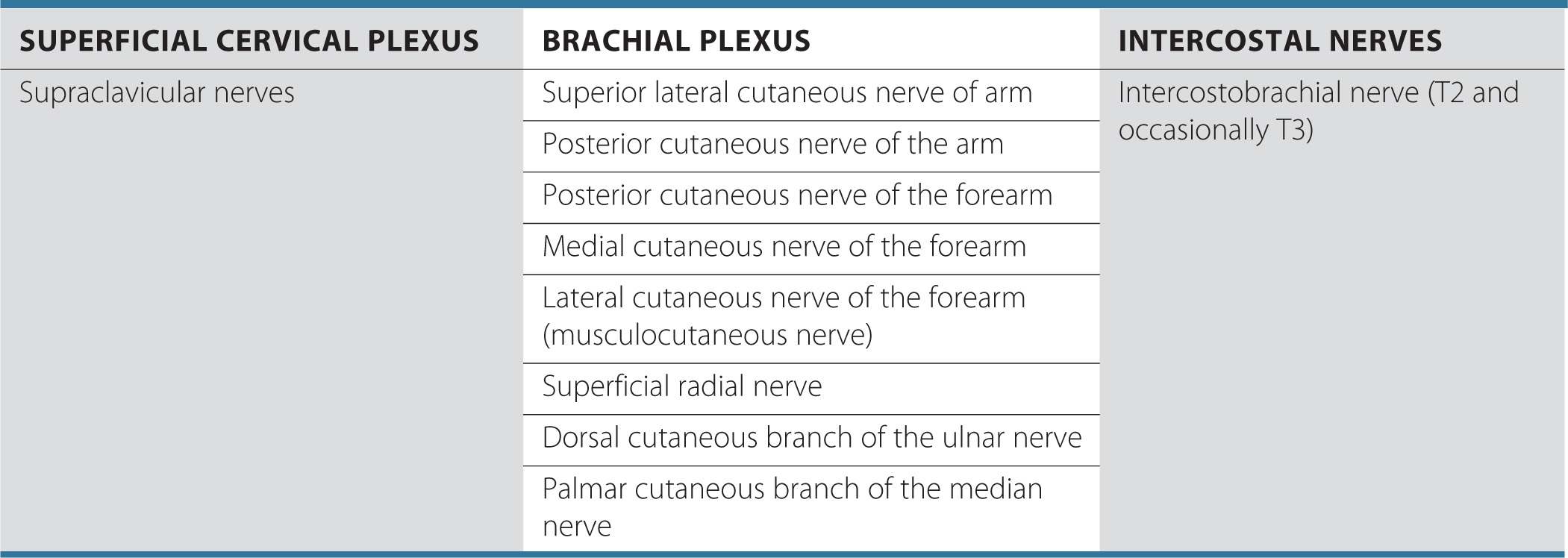
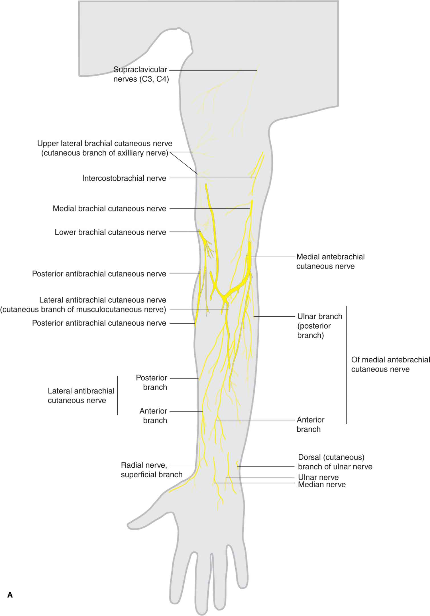
FIGURE 17-2. (A) Cutaneous innervation of the upper extremity (front).
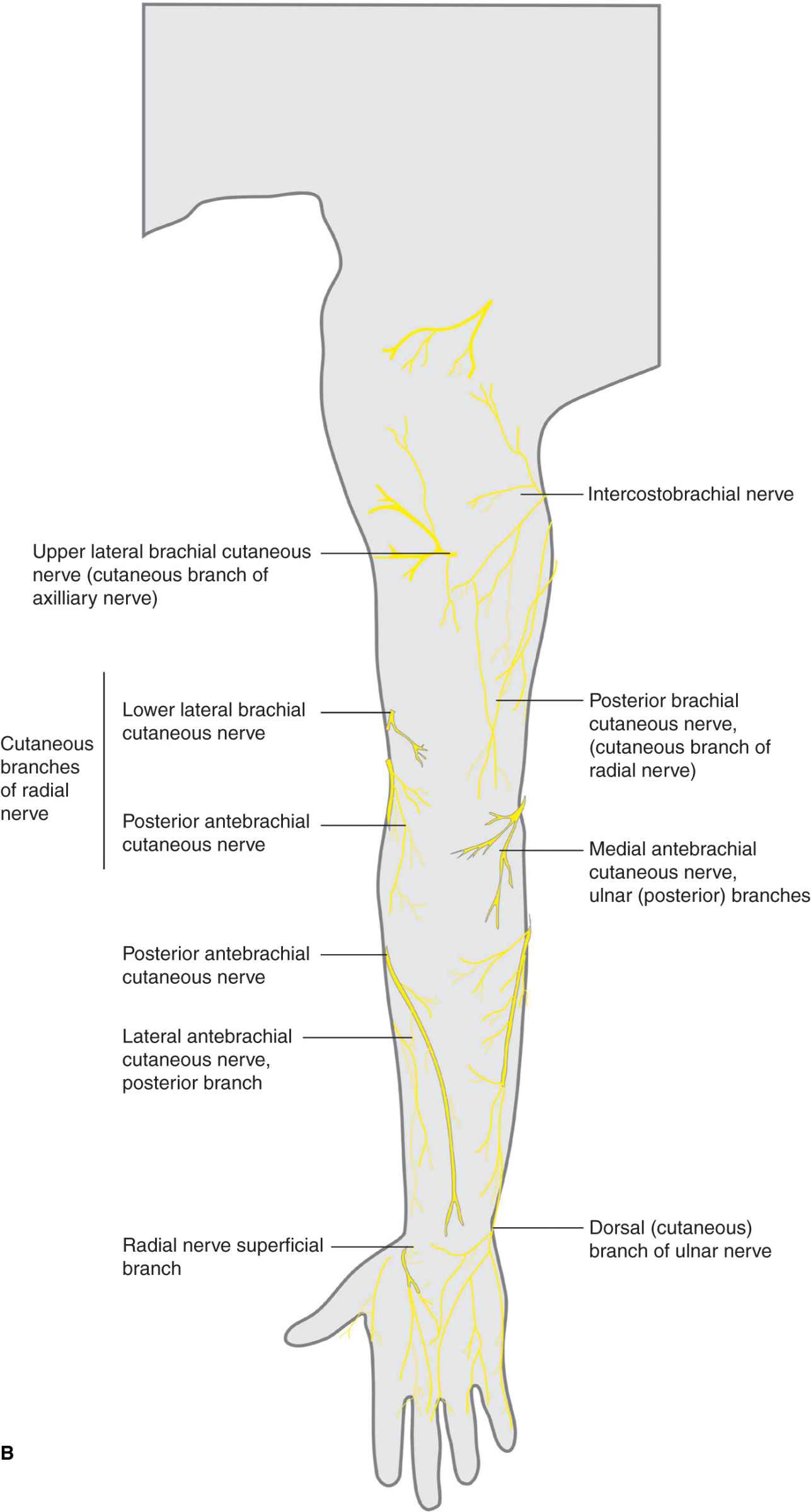
FIGURE 17-2. (B) Cutaneous innervation of the upper extremity (back).
Intercostobrachial Nerve Block
Anatomy
The intercostobrachial nerve is the lateral cutaneous branch of the ventral primary ramus of T2. It provides innervation to the skin of the axilla and the medial aspect of the proximal arm. The intercostobrachial nerve communicates with the medial cutaneous nerve of the arm, which is a branch of the brachial plexus (Figure 17-3). Both nerves are anesthetized by subcutaneous infiltration of the skin of the medial aspect of the arm.
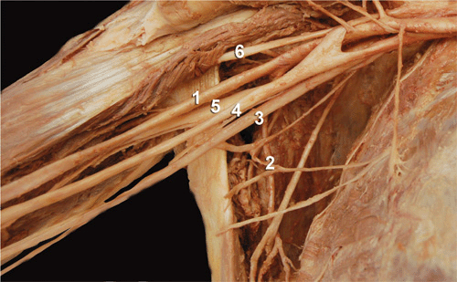
FIGURE 17-3
Related posts:


Stay updated, free articles. Join our Telegram channel

Full access? Get Clinical Tree


