Introduction
Maternal cardiorespiratory arrest is fortunately an infrequent yet catastrophic event. Triennial audits from the UK have repeatedly confirmed that major haemorrhage, thromboembolism, amniotic fluid embolism and hypertensive disease of pregnancy (PET and eclampsia) account for the majority of ‘direct’ obstetric related deaths. Data from the latest Centre for Maternal and Child Enquiry (CMACE) triennial report (2011) indicate a maternal mortality rate in the UK of 14/100,000; sepsis is now an additional leading ‘direct’ cause of death. Cardiac disease continues to be the leading ‘indirect’ cause of death, which is attributed to the increased prevalence of lifestyle risk factors (obesity, smoking and increased maternal age), peripartum cardiomyopathy, aortic dissection and the increased number of women surviving and becoming pregnant with pre-existing complex congenital heart disease.
Maternal collapse (which may or may not lead to cardiorespiratory arrest) occurs in 3–6 mothers per 1000 pregnancies. Major haemorrhage is overwhelmingly the most likely cause and increasingly better recognition and management has significantly reduced mortality in this situation.
Whether maternal collapse or cardiorespiratory arrest, this is a stressful and emotive situation in which there are potentially two lives that may be lost. The best hope for fetal survival is maternal survival.
Physiological changes
The physiological changes that occur during advanced pregnancy may alter management of cardiorespiratory arrest in the parturient: the ABC approach requires some modifications.
Airway
Laryngeal and tracheal oedema is more prevalent in advanced pregnancy and the pharyngeal mucosa is hyperaemic and more friable. The cross-sectional area of the upper airway is narrower in pregnancy. There is an increasing risk of aspiration from progesterone-induced gastro-oesophageal reflux and use of cricoid pressure may be necessary. Difficulty with tracheal intubation is more common in late pregnancy. Some of the standard indicators of a potentially difficult airway (e.g. short thyromental distance and poor jaw slide) may improve as pregnancy advances; thus, these surrogate markers are unreliable for predicting difficult tracheal intubation. The difficulties associated with tracheal intubation may also relate to:
- Large breasts
- Full dentition
- Glottic oedema
- Poorly applied cricoid pressure
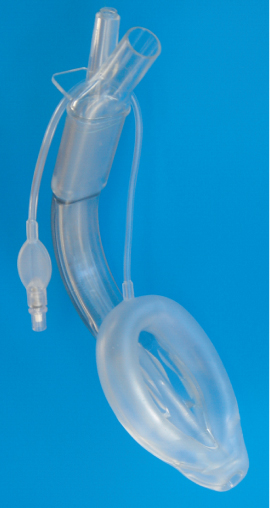
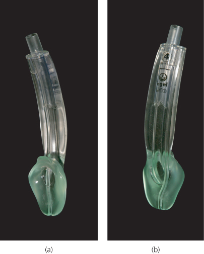
Potentially difficult tracheal intubation and the threat of aspiration are reasons for the individual who is not skilled in tracheal intubation to use a supraglottic airway device with a gastric drain port for airway control during CPR. Such devices include the LMA Supreme and the igel airway (Figures 15.1 and 15.2).
Breathing
The gravid uterus displaces the diaphragm causing a decrease in functional residual capacity (FRC) and decrease in chest wall compliance. This makes ventilation of the lungs more difficult and may necessitate higher inflation pressures during positive pressure ventilation—optimal bag-mask ventilation is impaired. Intrapulmonary shunting increases in pregnancy (up to 15%) and together with the increased oxygen requirements during advanced pregnancy, contributes to rapidly developing hypoxia during apnoea.
Maternal arterial carbon dioxide values are lower than normal because of hyperventilation caused by a progesterone-driven increase in tidal volume and minute ventilation. This may reduce the buffering capacity of the plasma and makes the development of an acidosis more likely in the setting of cardiorespiratory arrest.
Circulation
Plasma volume is increased in the parturient and there is a physiological or dilutional anaemia. This reduces oxygen carrying capacity. The heart rate increases and cardiac output may be up to 50% greater in the advanced stages of pregnancy. There is a decrease in systemic vascular resistance, which lowers systolic blood pressure. Uterine blood flow may be as much as 12% of cardiac output and this increases the risk of massive haemorrhage from this source.
The circulatory adjustments during CPR focus on avoidance of aortocaval compression. This phenomenon reduces both blood supply to the uterus (and therefore fetus) and, more importantly, maternal venous return to the heart. Cardiac output may be reduced by up to 35–40% in the supine parturient. Furthermore, maternal cardiac output may be persistently impaired despite efforts to reduce compression of the inferior vena cava (IVC) until delivery of the foetus. The risk for IVC compression is greater as pregnancy progresses and begins to occur as the uterus becomes visible above the pelvis. The problem is compounded by multiple pregnancy and conditions that cause pathological uterine enlargement (e.g. polyhydramnios).
Table 15.1 shows the physiological changes in advanced pregnancy and the potential effect on CPR.
Table 15.1 Physiological changes in advanced pregnancy and the potential effect on CPR.
| Change in pregnancy | Potential impact during CPR | |
| Cardiovascular system | ||
| Plasma volume | Increased by up to 50% | Dilutional anaemia Reduced oxygen carrying capacity |
| Heart rate | Increased by up to 30% | Increased oxygen and CPR demand |
| Cardiac output | Increased by up to 50% | Increased oxygen and CPR demand |
| Systemic vascular resistance | Decreased by up to 40% | Reduced coronary and cerebral perfusion during CPR |
| Venous return | Decreased by pressure of gravid uterus on IVC | Significant reduction in cardiac output generated by CPR |
| Uterine blood flow | Up to 12% of cardiac output | Potential for rapid and massive haemorrhage |
| Respiratory system | ||
| Tidal volume and minute ventilation | Increased by up to 45% | Hyperventilation reduces arterial PCO2 and plasma HCO3 Reduced buffering capacity |
| Oxygen consumption | Increased by up to 30% | Rapid development of hypoxia |
| Functional Residual Capacity | Decreased by up to 25% by pressure of gravid uterus | Increased intrapulmonary shunting. |
| Chest wall compliance | Reduced | Higher airway pressures Difficulty with ventilation |
| Pharynx | Hyperaemic | Airway narrowing Difficult tracheal intubation Bleeding if damaged |
| Larynx | Hyperaemic and oedema | Airway narrowing Difficult tracheal intubation |
| Other changes | ||
| Gastric motility | Reduced | Increased risk of aspiration |
| Lower oesophageal sphincter | Relaxed | Increased risk of aspiration |
| Uterus | Enlarged Up to 10% of cardiac output | Reduction in venous return from IVC compression Diaphragm splinting reduces lung compliance and increases intrapulmonary shunting Potential for massive haemorrhage |
| Weight | Increased | Transfer more difficult Increased breast size may impair ventilation, interfere with tracheal intubation and make positioning of defibrillator pads more difficult |
CPR, cardiopulmonary resuscitation; IVC, inferior vena cava.
Cardiopulmonary resuscitation guidelines 2010
The 2010 resuscitation guidelines for adults are followed with a few modifications for maternal resuscitation. Standard drug doses, defibrillation energies and compression ventilation ratios are used.
Drugs
The dose of adrenaline for all cardiac arrests and the dose of amiodarone for shock resistant ventricular fibrillation remain 1 mg and 300 mg respectively. Fibrinolytic drugs are recommended for massive pulmonary embolism although subsequent Caesarean section will be challenging. The potential effects on the fetus should not preclude their use during cardiac arrest.
The gravid uterus may reduce venous return—deliver drugs via a route that avoids the IVC, such as the upper limb. If venous access is difficult, insert an intra-osseous needle in the upper humerus.
Defibrillation
Transthoracic impedance in pregnancy is the same as the non-pregnant state; the same defibrillation energies are used. Delivery of an electric shock across the maternal chest does not harm the fetus. Current travelling directly through the uterus, for example electrocution from domestic electricity, may injure the fetus. Use of self-adhesive, multifunction electrodes/pads eliminates placement problems associated with large breasts and lateral tilt.
Chest compression
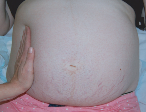
The 2010 guidelines emphasise improvement of the quality of chest compressions by increasing both the depth (5–6 cm) and frequency (100–120 per minute), ensuring adequate chest wall recoil and minimises interruption to chest compressions. Aortocaval compression must be avoided during CPR in advanced pregnancy. Although manikin studies have demonstrated that it is possible to perform chest compressions with a patient laterally tilted at various angles, in comparison with the supine position the maximum compression force achieved is reduced significantly. Therefore chest compressions are performed initially with the mother fully supine and the uterus manually displaced laterally. This relieves aortocaval compression while enabling excellent quality chest compressions (Figures 15.3 and 15.4).
< div class='tao-gold-member'>