Causes of cardiac arrest in pregnancy
Obstetric
Non-obstetric
Iatrogenic
Eclampsia
Pulmonary embolism
General anesthesia-induced
Amniotic fluid embolism
Sepsis
Regional anesthesia-induced
Postpartum hemorrhage
Trauma
Other medications (e.g., magnesium)
Antepartum hemorrhage
Myocardial infarction
Anaphylaxis
Peripartum cardiomyopathy
Aortic dissection
Status asthmaticus
Anaphylaxis
Amniotic fluid embolism, an etiology unique to the pregnant patient, occurs in 1 in 40,000 pregnancies [6]. It is accountable for 10% of peripartum mortality, and maternal mortality rate varies between 20 and 60% [6, 7]. The exact pathophysiology of amniotic fluid embolism is unclear, but current theories are based on the overwhelming maternal inflammatory response to fetal tissue. Initially, disruption of the maternal-fetal barrier, which can occur even in routine labor, leads to release of fetal tissue into maternal circulation. This triggers a brusque maternal inflammatory response, similar to the inflammatory response in anaphylaxis. Indeed, the term “anaphylactoid syndrome of pregnancy” is often used in the literature to describe the condition. Next, a period of uterine hypertonicity leads to fetal distress. Often, it is the signs of fetal distress including late decelerations and fetal bradycardia that are first recognized by providers before maternal signs and symptoms. A consumptive coagulopathy follows, similar to disseminated intravascular coagulopathy.
Amniotic fluid embolism can present as sudden maternal dyspnea and oxygen desaturation during labor or shortly following delivery. Cardiovascular collapse ensues and cardiac arrest is common. Women who survive initial cardiac arrest are prone to acute lung injury and acute respiratory distress syndrome, in addition to anoxic brain injury. Fetal distress occurs along with maternal distress (and may even precede maternal symptoms) as blood is shunted from the uterus to the maternal central circulation for preservation of cardiac and neurologic function.
Treatment for amniotic fluid embolism is primarily supportive. Anoxic brain injury may occur quickly, so a definitive airway should be established early in the resuscitation, and patients should be treated with 100% oxygen [8]. Intravascular support in the way of intravenous fluids, blood products, and vasopressors, if necessary, is recommended. Blood products should be considered before clinical signs of hemorrhagic shock. The use of specific factors such as factor VII is not recommended, as these have been linked to vaso-occlusive events [6].
Non-obstetric Causes
Non-obstetric causes of cardiac arrest in pregnancy include sepsis, trauma, myocardial infarction, aortic dissection, anaphylaxis, status asthmaticus, and pulmonary embolism (PE). The most common cause of cardiac arrest in pregnancy in the United States is PE [9]. PE complicates 1 in 1,000 pregnancies, and pregnant women are up to ten times more likely to have a venous thromboembolism than nonpregnant women [10, 11]. When PE is the cause of cardiac arrest, standard advanced cardiac life support (ACLS) therapies are less likely to be effective, necessitating early recognition of this pathology [12]. Clues to PE as the etiology of cardiac arrest include pulseless electrical activity as the initial rhythm as well as a dilated right ventricle on bedside ultrasound [13, 14]. Treatment includes standard ACLS protocol and fibrinolytic therapy. Fibrinolytic options include recombinant tissue plasminogen activators, or synthetic derivatives such as streptokinase and urokinase [15]. A recent meta-analysis of hemodynamically unstable pregnant patients with PE showed the risk of major bleeding following fibrinolysis was low, and the fetal demise rate was only 2.6% [16]. There have been multiple case reports of successful use of fibrinolytic therapy in unstable pregnant women with massive PE, including patients at 26 weeks and 32 weeks with good outcomes [17–19]. Another somewhat novel therapeutic option is extracorporeal membrane oxygenation (ECMO). There have been several case reports showing the use of ECMO for PE with favorable outcomes for both mother and fetus [20, 21].
Iatrogenic Causes
Iatrogenic causes of maternal cardiac arrest include anesthesia-related arrest (both general and regional anesthesia) and medication induced, specifically magnesium toxicity. Anesthesia-induced cardiac arrest is the seventh leading cause of maternal death in the United States [22], although mortality rates are declining [23]. The decline in maternal deaths related to general anesthesia has been attributed to increase the use of pulse oximetry and capnography and utilization of difficult intubation algorithms including the use of supraglottic airway devices. The reduction in maternal deaths from local anesthesia is likely due to using a lower concentration of anesthetics, an increase in general awareness regarding anesthetic toxicity, and an overall emphasis on safety practices [22].
Pregnant patients with systemic anesthetic toxicity present with respiratory and cardiovascular depression, which can eventually lead to complete cardiopulmonary collapse. Treatment is supportive and centered on early recognition, fluid resuscitation, and airway management. Prevention of hypoxia and metabolic acidosis is critical [22]. For patients with complete cardiopulmonary collapse refractory to standard resuscitative measures, lipid emulsion therapy can be considered, though a recent review was inconclusive regarding the efficacy of lipid emulsion therapy for systemic anesthetic toxicity [24].
Another cause of iatrogenic cardiac arrest in pregnancy is magnesium toxicity. Magnesium is a standard therapy for patients with preeclampsia and eclampsia, and there is a risk of cardiopulmonary collapse if toxic levels are reached [25]. Fortunately, toxicity is now rare, as many hospitals have protocols in place to monitor for overdose; however, the possibility still exists and was addressed in recent guidelines from the American Heart Association (AHA) on cardiac arrest [26]. Toxic levels are reached at levels greater than 3.5 mmol/L, although the exact dosing to reach this level is unclear [27]. Symptoms of magnesium toxicity include loss of deep tendon reflexes, respiratory depression, somnolence, and cardiac arrest. If magnesium toxicity is suspected, the infusion should be stopped immediately, and 10 mL of 10% calcium gluconate can be administered [28, 29].
Resuscitation Considerations
ACLS algorithms are generally the same in pregnancy including defibrillation doses and medical management. However, three important differences exist when resuscitating a pregnant patient: (1) positioning of the patient, (2) airway management and anticipation of a difficult airway, and (3) early consideration of perimortem cesarean delivery (PMCD). Additionally, intravenous access should be obtained above the diaphragm in pregnant patients.
Positioning of the Patient
Positioning of a pregnant patient in cardiac arrest is one of the key differences from resuscitating the nonpregnant patient. Once the uterus is palpable above the umbilicus (around 20 weeks), the uterus compresses the aorta and vena cava and decreases venous return when the woman lies supine (aortocaval compression). Chest compressions only produce 30% of normal cardiac output in healthy, nonpregnant patients [30]. In pregnant patients, cardiac output is reduced by an additional 60% in the supine position due to aortocaval compression. Therefore, in pregnant patients, chest compressions in the supine position produce 10% of normal cardiac output at best [31].
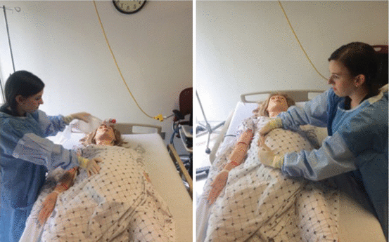
Due to aortocaval compression and the resultant limitations on cardiac output, positioning of the pregnant patient is crucial. There are two methods that have been historically used to help decrease aortocaval compression: manual left uterine displacement and lateral tilt. Manual left uterine displacement is the technique currently recommended by the AHA [32]. This can be done using a one-handed technique from the right side of the bed, or with two hands from the left side of the bed (Fig. 10.1a, b). The two-handed approach should be a “pulling” motion instead of a “pushing” motion. Lateral tilt has also been used, although this requires a dedicated foam or wooden wedge and may be less effective [33].


Fig. 10.1
Demonstration of manual uterine displacement on a simulated gravid patient. One-handed approach from the patient’s right side, and two-handed approach from patient’s left. Note the “pulling” motion of the provider, as opposed to a “pushing” maneuver when using the two-handed approach
The AHA recommends chest compressions be performed without modifications for the pregnant patient. This includes compressions performed over the lower half of the sternum at a rate of greater than 100 compressions per minute, compressing at a depth of at least 2 in. (5 cm), allowing for complete chest recoil. The compressions to ventilations ratio remains 30 to 2.
Airway Management
Airway management of obstetric patients can be challenging due to anatomical and physiologic changes related to pregnancy. Physiologic changes such as increases in breast tissue and overall weight gain decrease chest wall compliance, making bag-valve-mask ventilation difficult [34]. During late pregnancy, increases in intra-abdominal pressure, a laxity of the lower esophageal sphincter, and slowed gastric emptying increase aspiration risks during endotracheal intubation. In pregnancy, there is an overall increased oxygen demand and decreased functional residual capacity leading to increased susceptibility to hypoxia and rapid desaturation. One study by McClelland et al. in 2009 demonstrated this rapid desaturation during induction of anesthesia, with pregnant subjects reaching SpO2 less than 90% within 5 minutes compared to 9 minutes in nonpregnant subjects. Obesity and active labor accelerate desaturation times, with pregnant patients often becoming significantly hypoxic in less than 3 minutes [35].
Anatomic changes in pregnancy include an overall increase in laryngeal edema. Airway tissue becomes edematous and may prevent the passage of larger (size 7.0 or greater) endotracheal tubes. The Mallampati classification system, historically utilized to evaluate a patient’s ability to open their mouth for endotracheal intubation, has been shown to advance one to two classes during pregnancy. These airway changes can persist up to 2–3 weeks following delivery [31].
For all these reasons, emergency physicians should anticipate obstetric patients to be difficult to intubate. Difficult or failed intubation is a major contributory factor to poor maternal outcomes following cardiac arrest [36]. Physicians should consider selecting smaller-sized endotracheal tubes and limit the number of tube passes, as multiple attempts can worsen pre-existing edema. Endotracheal intubation is preferred over supraglottic airways, but laryngeal mask airways (LMAs) and similar adjuncts should be considered following two unsuccessful intubation attempts [32].
Perimortem Cesarean Delivery
Perimortem cesarean delivery (PMCD) describes the procedure of delivering a fetus during maternal cardiac arrest [37]. Historically, the procedure has been performed to potentially benefit the fetus [31]. Over the years, however, it had been observed that when cardiopulmonary resuscitation (CPR) was performed on pregnant patients, return of spontaneous circulation (ROSC) was often achieved after “emptying the uterus” and relieving aortocaval compression [37]. A paradigm shift has since occurred: once considered purely fetocentric, PMCD is now recommended as a primarily maternal resuscitative measure. Indeed, some authors have replaced the term “perimortem cesarean delivery” with “resuscitative hysterotomy” in the literature.
As a general rule, the fundus of the uterus reaches the umbilicus around 20 weeks gestation. It is at this point that the uterus begins to cause significant compression of both the aorta and vena cava, impacting venous return to the maternal heart and increasing maternal afterload [31]. Therefore, any pregnant patient in cardiac arrest with a palpable uterus at, or above, the level of the umbilicus is a candidate for PMCD [4]. Performing the procedure on patients less than 20 weeks gestation is not recommended, as it is not likely to benefit the mother or the fetus.
Anoxic brain injury occurs after 4–6 min of cessation of blood flow. [37] Fetal neurologic sequelae tend to occur later than in the mother following an anoxic event [38]. Historically, PMCD was advised after 4 min of CPR without return of ROSC with the goal of having the baby delivered within 1 minute (the “4-minute rule”). However, this standard can be difficult to attain. A retrospective case review by Benson et al. found 90% of perimortem cesarean deliveries occurred after the 5-minute mark [39]. Additionally, multiple studies have shown positive maternal and fetal outcomes following deliveries well outside the 4-minute window [4, 31, 40]. Some authors recommend performing the procedure immediately in patients with non-shockable rhythms (pulseless electrical activity and asystole) [37]. In patients with ventricular dysrhythmias (“shockable rhythms”), 4 minutes of ACLS prior to PMCD is still advised [41].
Contraindications to PMCD include gestational age less than 20 weeks (or a uterine fundus below the level of the umbilicus in multiple pregnancies), maternal ROSC, and prolonged maternal arrest (typically greater than 15 minutes, although fetal survival has been described in the literature as long as 45 minutes after maternal arrest [42]). Little equipment is needed, and the procedure can be performed with only a scalpel if necessary (Table 10.2). The procedure should be performed by the provider with the most surgical experience, and resuscitation teams for both the mother and fetus should be present.
Table 10.2


Perimortem cesarean section procedure, adapted from Parry R et al. [41]
Stay updated, free articles. Join our Telegram channel

Full access? Get Clinical Tree


