HELPERR
H
Call for help
E
Evaluate for episiotomy
L
Legs (McRobert’s Maneuver by assistant)
P
Suprapubic pressure (by assistant)
E
Enter maneuvers/internal rotation (Rubin’s, Woods Corkscrew)
R
Remove posterior arm
R
Roll the patient (Gaskin Manuever)
ALARMER
A
Ask for help
L
Leg hyperflexion (McRoberts)
A
Anterior shoulder disimpaction (suprapubic pressure)
R
Rotational maneuvers (Rubin II, Woods Corkscrew)
M
Manual delivery of posterior arm
E
Evaluate for episiotomy
R
Roll the patient (Gaskin)
The first step in approaching a shoulder dystocia is to call for help. Even more assistance is often needed than would be required in a typical precipitous delivery. This will become important in the various maneuvers described. There is also greater potential for fetal injury and need for fetal resuscitation.
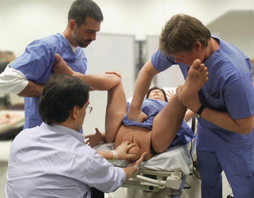
The first or primary maneuvers in a shoulder dystocia are McRoberts maneuver and suprapubic pressure. These have the highest chance of success and the lowest risk of fetal injury. These two maneuvers move the maternal pubic symphysis more cephalad and typically release the anterior shoulder. McRoberts maneuver requires two assistants—each one holding a leg with the knees and hips flexed and the thighs held against the abdomen (Fig. 7.1). McRoberts maneuver has been shown by x-ray not to increase the pelvic diameter but rather to facilitate cephalad rotation of the symphysis pubis with flattening/straightening of the sacrum [27]. This allows the posterior shoulder to be pushed over the sacral promontory and fall into the hollow of the sacrum, while the symphysis rotates over the impacted anterior shoulder.


Fig. 7.1
McRoberts maneuver performed with two assistants. Assistant on patient’s left applies suprapubic pressure. Image courtesy CAE Healthcare, with permission
One assistant should apply suprapubic pressure with the heel of their hand just over the mother’s pubic bone and over the anterior fetal shoulder. Downward and lateral pressure applied to the posterior aspect of the impacted, anterior fetal shoulder should facilitate adduction of the anterior shoulder, decreasing the fetal diameter and allowing for passage of the anterior shoulder under the symphysis pubis. Initially continuous, the pressure can also be applied in a rocking motion similar to chest compressions. Fundal pressure should never be applied as it only worsens impaction on the pelvic brim, potentially injuring the fetus or mother [28]. McRoberts alone has been shown to be successful between 40 and 58% of the time [22, 29, 30]. When suprapubic pressure is added, success rates of 54 and 58% have been found [22, 29].
If primary maneuvers (McRoberts and suprapubic pressure) are unsuccessful, secondary maneuvers to consider include rotational maneuvers or delivery of the posterior arm. These are necessary in approximately 1/3 of cases [31]. Although typically signifying a greater degree of dystocia, these maneuvers are not actually associated with a higher risk of maternal or fetal injury [19, 32]. An episiotomy should be considered, but not routinely performed when a shoulder dystocia is encountered. An episiotomy will not in itself alleviate the primary problem in a shoulder dystocia (a bone on bone impaction), and prophylactic episiotomy with delivery does not reduce the risk of shoulder dystocia occurring [33]. Episiotomy has also been associated with a sevenfold increase in the rate of severe perineal trauma [34]. However, episiotomy does provide additional space for the physician’s hand to successfully perform the various internal maneuvers to alleviate the shoulder dystocia. With the high success rate of McRoberts maneuver and suprapubic pressure, it is most appropriate to wait until additional maneuvers are required before performing an episiotomy.
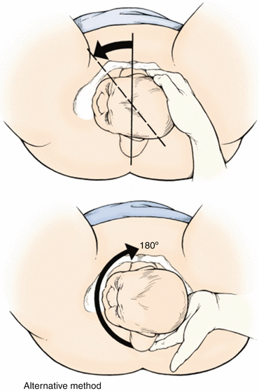
The focus of internal rotation maneuvers is to allow entry of the fetal shoulders into the pelvic inlet in a transverse or oblique diameter. The most popular internal rotation maneuvers include the Rubin II maneuver and Woods corkscrew maneuver. Rubin II maneuver involves placement of fingers behind the posterior aspect of the anterior shoulder of the fetus and rotating the shoulder toward the fetal chest (Fig. 7.2). The resulting adduction of the fetal shoulder girdle reduces the shoulders’ diameter, potentially alleviating the dystocia. Woods corkscrew maneuver involves continued application of the pressure on the anterior shoulder (as in Rubin II), with additional upward pressure placed on the anterior aspect of the fetal posterior shoulder toward the fetal back, leading to posterior shoulder abduction (Fig. 7.2). This creates additive force in the same circumferential arc as Rubin II, resulting in more effective rotation. The McRoberts maneuver also can be applied during this maneuver and may facilitate its success. If the Rubin II or Woods corkscrew maneuvers fail, the reverse Woods corkscrew maneuver may be tried. In this maneuver, the physician’s fingers are placed on the back of the posterior shoulder of the fetus, and the fetus is rotated in the opposite direction as in the Woods corkscrew or Rubin II maneuvers.


Fig. 7.2
Rubin II rotation of the anterior shoulder forward into the fetal chest (top). Reverse Woods corkscrew place fingers behind posterior shoulder and rotate fetus in the opposite direction of Rubin II (bottom). From Gabbe SG, eds. Obstetrics: Normal and Problem Pregnancies. Philadelphia: Churchill Livingstone, 2017; with permission
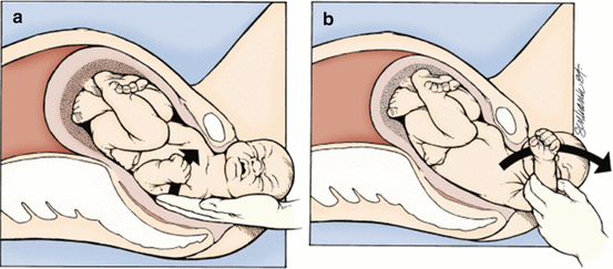
If internal rotation maneuvers fail, delivery of the posterior arm should be considered. Place a hand behind the posterior fetal shoulder, tracing the arm to the elbow, where pressure is applied to the antecubital fossa to flex the fetal forearm (Fig. 7.3). The arm is then swept out over the fetal chest and delivered. Often, the fetus follows with spontaneous rotation after the arm is removed, leading to alleviation of impaction of the anterior shoulder. Grasping or pulling of the arm or pressure directly on the humeral shaft should be avoided. This maneuver is associated with an increased rate of humeral fracture.


Fig. 7.3
Delivery of the posterior arm. The operator here inserts a hand (a) and sweeps the posterior arm across the chest and over the perineum (b). Care should be taken to distribute the pressure evenly across the humerus to avoid unnecessary fracture. From Gabbe SG, eds. Obstetrics: Normal and Problem Pregnancies. Philadelphia: Churchill Livingstone, 2017; with permission
Rotational maneuvers were not associated with a higher fetal injury risk compared with McRoberts maneuver [35]. Individual maneuvers (Rubin, Woods screw, delivery of posterior arm) were not associated with increased composite morbidity, neonatal injury, or neonatal depression after adjusting for nulliparity and duration of shoulder dystocia.
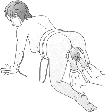
The next maneuver to consider is to roll the patient into an “all fours” position—referred to as the Gaskin maneuver (Fig. 7.4). This has been demonstrated to increase obstetric conjugate by as much as 10 mm with pelvic outlet increasing by as much as 10 mm [36]. This increased space with the addition of gravity is often enough to deliver the posterior shoulder and arm and allow for disimpaction of the anterior shoulder from the pubic symphysis. Gaskin maneuver was found to be successful in over 80% of women who required progression to this maneuver [37]. Internal maneuvers may be repeated while the patient is on “all fours” if simply moving into this position does not result in delivery.


Fig. 7.4
The Gaskin position. The “all fours” position exploits the effects of gravity and increased space in the hollow of the sacrum to facilitate delivery of the posterior shoulder and arm. From Gottlieb AG, Galan HL. Shoulder dystocia: An update. Obstet Gynecol Clin N Am. 2007;34:501–531; with permission
Heroic maneuvers should be considered only after all other maneuvers have failed. These include cephalic replacement (Zavanelli) followed by cesarean section, symphysiotomy (used more widely in developing countries), deliberate clavicular fracture (to reduce bisacromial diameter), hysterotomy (to allow rotation from intrauterine pressure), and delivery under general anesthesia.
Management of Umbilical Cord Prolapse
Umbilical cord prolapse occurs when all or part of the umbilical cord precedes the fetal presenting part. This is associated with abnormal presentations, situations where the presenting part does not fill the entire cervical canal, or with a long umbilical cord. Resulting compression of the umbilical cord can result in fetal hypoxia, distress, and asphyxiation.
With an umbilical cord prolapse and a viable fetus, cesarean delivery is the method of delivery of choice. Maneuvers to preserve circulation should be instituted immediately. Attempt should be made to digitally elevate the presenting part from the umbilical cord and place the mother in Trendelenburg or in an “all fours” position. Advise the patient not to push. Placement of a Foley catheter and instilling 500 mL saline may help lift the fetus off the cord. Preparation for emergency cesarean should be underway. Manipulation and cord trauma should be kept to a minimum. If there is no option for surgical management, the umbilical cord may be pushed gently in a retrograde fashion above the presenting part. Subsequent to delivery, the physician should be prepared for resuscitation of a distressed infant.
Management of Breech Delivery
Breech presentation is the presentation of fetal feet or buttocks. It is an obstetric emergency that occurs in 3–4% of term pregnancies [38, 39] with a higher incidence in preterm deliveries (due to the fact that final natural fetal rotation may not yet have occurred) and with advanced maternal age [40]. Breech presentation is associated with a morbidity rate 3–4 times greater than that of normal cephalic presentations [38]. It increases the likelihood of umbilical cord prolapse and dystocia during delivery. During a normal delivery, the head, which is larger than the buttocks, maximally dilates the cervix with the body following, while in a breech delivery, the buttock does not adequately dilate the cervix to allow for passage of the diameter of the head. A dystocia can then occur after delivery of the buttock and body as the head is stuck with resultant umbilical cord compression, fractures, spinal cord injuries, and potential asphyxiation and fetal death as delivery of the head is attempted.
Stay updated, free articles. Join our Telegram channel

Full access? Get Clinical Tree


