, Corinna Eleni Psomadakis2 and Bobby Buka3
(1)
Department of Family Medicine, Mount Sinai School of Medicine Attending Mount Sinai Doctors/Beth Israel Medical Group-Williamsburg, Brooklyn, NY, USA
(2)
School of Medicine Imperial College London, London, UK
(3)
Department of Dermatology, Mount Sinai School of Medicine, New York, NY, USA
Keywords
Blistering dactylitisBullousBullaBullaeBacterial infectionBlister Streptococcus Staphylococcus BitesMRSAAntibioticsRabiesTetanus
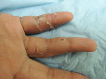
Fig. 12.1
Partial thickness shedding of digital skin with large collarets of scale

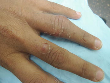
Fig. 12.2
Scale collarets are oftentimes an indication of an infectious process
Primary Care Visit Report
A 37-year-old female with no past medical history presented with redness and peeling skin on bilateral hands and fingers after sustaining a dog bite 2 weeks prior. The patient was bitten on both hands when she jumped in to separate a dog fight. The patient initially went to an emergency room and was given tetanus and rabies vaccines . Three days later, her fingers were swelling so she went back to the ER and was admitted for 3 days of IV antibiotics (vancomycin and ampicillin/sulbactam) and was discharged on amoxicillin/clavulanate and doxycycline, which she was taking at the time of this visit.
While the pain and stiffness in her fingers had improved somewhat, the patient remained concerned because she noticed the night prior that certain areas had become “red and bumpy” and about 4 h later, the skin on her fingers began to peel. The patient had no fever or chills, and felt fine otherwise.
At the time of this visit, the patient was status post three rabies vaccinations on Day 0, Day 3 and Day 7 and was due for the fourth vaccine, though she found out from its owner that the dog who bit her was up to date on its rabies vaccinations .
Vitals were normal. On exam, on bilateral hands, there were multiple erythematous lesions with erythematous bases at the sites of the dog bites . There was peeling on the periphery of many of the lesions, and many were indurated and tender to palpation.
Specifically, the right hand pinky finger had erythema and tenderness to palpation spanning the full circumference of her finger (dorsal and ventral aspect) at the distal-interphalangeal joint and extending proximally with limited active range of motion (but full passive range of motion) at the distal-interphalangeal joint. At the periphery of the erythema, the skin was peeling. The left thumb had a 1.5 cm × 1.0 cm erythematous lesion with peeling skin at the periphery, with some induration and tenderness to palpation. The left fourth finger had a 3.0 cm × 1.5 cm erythematous, tender and indurated lesion with peeling at the periphery at the proximal-interphalangeal joint. She had limited active range of motion at this joint (normal passive range of motion). No warmth on any of the lesions. Peripheral pulses were normal.
Since the patient was already on antibiotics yet continued to have further worsening of pain and developed new areas of erythema and peeling, I was concerned about an abscess and referred her immediately to a hand surgeon for further evaluation. The hand surgeon examined her and was not concerned for an abscess but did recommend her for physical therapy.
Stay updated, free articles. Join our Telegram channel

Full access? Get Clinical Tree


