Anesthesia for Thoracic Surgery
Lung cancer has long been the most common cause of cancer mortality in the United States in men and has surpassed breast cancer as the leading cause of cancer deaths in women (Eisenkraft JF, Cohen E, Neustein SM. Anesthesia for thoracic surgery. In: Barash PG, Cullen BF, Stoelting RK, Cahalan MK, Ortega R, Stock MC, eds. Clinical Anesthesia. Philadelphia: Lippincott Williams & Wilkins; 2013:1030–1075). The increased incidence of lung cancer has led to an increase in the amount of noncardiac thoracic surgery performed in the United States.
I. Preoperative Evaluation
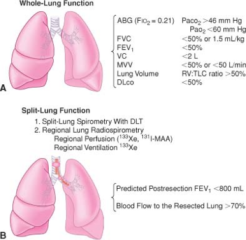
(Fig. 37-1). The preoperative evaluation of the patient for thoracic surgery should focus on the extent and severity of pulmonary disease and cardiovascular involvement. Thoracic surgery is known to be associated with high risk, and patient factors that have been associated with increased risk include advanced age, poor general health status, and chronic obstructive pulmonary disease (COPD).
History (Table 37-1)
Physical Examination (Table 37-2)
Electrocardiogram A patient with COPD may present with electrocardiographic features of right atrial and ventricular hypertrophy and strain.
Chest Radiography. Hyperinflation and increased vascular markings are usually present with COPD. Tracheal or carinal shift should be noted. Review of a computed tomography (CT) study often provides more information about tumor size and location than the chest radiograph.
Arterial blood gases. A common finding in arterial blood gas analysis of patients with COPD is hypoventilation and CO2 retention.
“Blue bloaters” (chronic bronchitis) are cyanotic, hypercarbic, hypoxemic, and usually overweight. These patients may hypoventilate when given high oxygen concentrations to breathe because of a decreased hypoxic drive.
“Pink puffers” (patients with emphysema) are typically thin, dyspneic, and pink, with essentially normal arterial
blood gas values. They present with an increase in minute ventilation to maintain their normal PaCO2, which explains the increase in work of breathing and dyspnea.
Table 37-1 Patient Medical History that Should be Obtained Before Thoracic Surgery
Dyspnea: quantitate as to activity required to produce; presence may warn of the need for postoperative ventilation)
Cough (characteristics of sputum)
Cigarette smoking
Exercise tolerance: increased risk when unable to climb two flights of stairs
Risk factors for acute lung injury: alcohol abuse, high ventilatory pressures, excessive fluid administration
Table 37-2 Physical Examination that Should be Performed Beofre Thoracic Surgery
Respiratory System
Cyanosis
Clubbing
Breathing rate and pattern (distinguish between obstructive and restrictive disease)
Breath sounds (wet sounds vs. wheezing)
Cardiovascular System
Presence of pulmonary hypertension
Function of left side of the heart (patients with valvular or ischemic heart disease)
Pulmonary Function Testing and Evaluation for Lung Resectability. Goals in performing pulmonary function tests in a patient scheduled for lung resection include (1) identification of the patient at risk of increased postoperative morbidity and mortality, (2) identification of the patient who will need short- or long-term postoperative ventilatory support, and (3) evaluation of the beneficial effect and reversibility of airway obstruction with the use of bronchodilators.
Spirometry. A patient with an abnormal vital capacity has a 33% likelihood of complications and a 10% risk of postoperative mortality.
In the past, a forced expiratory volume in 1 second (FEV1) of <800 mL in a 70-kg man had been considered an absolute contraindication to lung resection. However, with the advent of thoracoscopic surgery and improved postoperative pain management, patients with smaller lung volumes are now successfully undergoing surgery.
The ratio of FEV1 to forced vital capacity (FVC) is useful in differentiating between restrictive (normal ratio) and obstructive pulmonary disease (ratio low).
A ratio of residual volume to total lung capacity of >50% is generally indicative of a high-risk patient for pulmonary resection.
Flow-volume loops display essentially the same information as a spirometer but are more convenient for measurement of specific flow rates (Fig. 37-2).
Patients with obstructive airways disease (asthma, bronchitis, emphysema) typically have grossly decreased FEV1/FVC ratios.
Patients with restrictive disease such as (pulmonary fibrosis, scoliosis) typically have decreased FVC with a relatively normal FEV1.
Significance of Bronchodilator Therapy. Pulmonary function tests are usually performed before and after bronchodilator therapy to assess the reversibility of the airways obstruction. A 15% improvement in pulmonary function tests may be considered a positive response to bronchodilator therapy and indicates that this therapy should be initiated before surgery.
Split-lung Function Tests. Regional lung function studies serve to predict the function of the lung tissue that would remain after lung resection. A whole (two)-lung test may fail to estimate whether the amount of postresection lung
tissue will allow the patient to function at a reasonable level of activity without disabling dyspnea or cor pulmonale.
Table 37-3 Conditions that Correlate with Postoperative Complicatoins
Infection: Treat with broad-spectrum antibiotics
Dehydration: Hydration decreases viscosity of secretions and facilitates their removal
Electrolyte imbalance
Wheezing: Delay elective surgery until effective treatment has been instituted; steroids, sympathomimetics, cromolyn, parasympatholytics
Obesity
Cigarette smoking: Carboxyhemoglobin decreases in 48 hours; decrease in sputum production in 2–3 months
Cor pulmonale
Malnutrition
Computed Tomography and Positron Emission Tomography Scans
The CT scan can delineate the size of the tumor and reveal if there is airway or cardiovascular compression.
Positron emission tomography (PET) may be more accurate than CT for mediastinal staging and can be used to further evaluate lesions that are seen on a CT scan.
Diffusing Capacity for Carbon Monoxide
The ability of the lung to perform gas exchange is reflected by the diffusing capacity for carbon monoxide. (A predicted postoperative diffusing capacity for carbon monoxide <40% is associated with increased risk.)
Predicted postoperative diffusing capacity percent is the strongest single predictor of risk of complications and mortality after lung resection.
Maximal oxygen consumption (VO2 max) is a predictor of postoperative complications (<10 mL/kg/min indicates very high risk for lung resection). The preoperative evaluation of the patient for lung resection is summarized in Figure 37-1.
II. Preoperative Preparation
The wide spectrum of physiologic changes that occur during thoracic surgery puts patients at great risk of developing postoperative complications (Table 37-3).
Table 37-4 Invasive Monitoring for Thoracic Surgery | ||||||
|---|---|---|---|---|---|---|
|
III. Intraoperative Monitoring (Table 37-4)
Pulse oximetry is especially valuable during thoracic surgery because hypoxemia may occur during one-lung ventilation (OLV).
Dysrhythmias occur commonly both during and after thoracic surgery, making the usual need for continuous ECG monitoring even more important.
IV. One-Lung Ventilation
Indications for OLV may be categorized as absolute and relative (Table 37-5).
Methods of Lung Separation
Stay updated, free articles. Join our Telegram channel

Full access? Get Clinical Tree


