96 Vertigo
• The majority of cases of vertigo arise from a peripheral source (e.g., cranial nerve VIII, vestibular structures) rather than a central source.
• The Dix-Hallpike test (also called the Nylan-Bárány test) can help confirm the diagnosis of benign paroxysmal positional vertigo.
• Other causes should always be considered because the signs and symptoms of central vertigo can mimic those of peripheral vertigo.
• Central vertigo should be suspected in patients with any abnormal neurologic findings.
Peripheral Vertigo
Epidemiology
General
Vertigo is diagnosed in roughly half of patients reporting dizziness. It is defined as the sensation of movement when none exists. About 80% of cases of vertigo are due to a peripheral lesion (pathology of the vestibular structures or cranial nerve VIII). Although peripheral vertigo is benign and not life-threatening, it may cause significant disability.
Benign Paroxysmal Positional Vertigo
The most common type of vertigo is benign paroxysmal positional vertigo (BPPV).1 It occurs in all age groups but becomes more common with aging. Women are affected twice as often as men.
Pathophysiology
Peripheral vertigo results from pathology involving the vestibular system or the eighth cranial nerve. The vestibular organs are responsible for equilibrium. The two main components—the three semicircular canals and the two otolith organs (utricle and saccule)—are interconnected and contain endolymph. The three semicircular canals—posterior (inferior), horizontal (lateral), and anterior (superior, vertical, dorsal)—are arranged at right angles to each other and allow detection of rotation via movement of endolymph. Otoliths (also known as otoconia), which are calcium carbonate particles, are found in the saclike utricle and provide information on head tilt.
When there is no disease and the head is held motionless, the bilateral vestibular systems fire at a tonic resting frequency. When the head rotates, there is increased firing from one semicircular canal and decreased firing from the others. The cerebral cortex interprets this information, synthesizes it with signals from the visual and proprioceptive systems, and translates it into the consciousness of movement. Vertigo results when the end-organs fire inappropriately at different frequencies, which causes unequal input to the brainstem and cerebral cortex.
Nystagmus is an involuntary rhythmic movement of the eyes to and fro that is often seen in patients with vertigo. Nystagmus is defined in terms of the direction of the fast-phase component. The factors listed in Table 96.1 should be noted in all patients with nystagmus. It should be kept in mind that on extremes of lateral gaze, several-beat nystagmus is a normal finding in 60% of patients.
Table 96.1 Characteristics of Nystagmus
Direction | Left or right |
---|---|
Axis | Upbeat or downbeat |
Nature | Rotary/torsional or vertical |
Duration | Seconds, minutes, or persistent |
Associated Factors | Spontaneous or positionalEffect of visual fixation |
Benign Paroxysmal Positional Vertigo
BPPV results from a mechanical defect in the inner ear. Canalolithiasis is the most accepted hypothesis. According to this premise, the culprits are free-floating otoliths that have become displaced from the utricle and inappropriately enter and activate the end-organs of the semicircular canals.2 With certain head movements, a clump of otoliths moves, causes the endolymph to move with resultant inappropriate displacement of the cupula. Movement of the cupula triggers neural firing, with consequential vertigo and nystagmus. Less commonly, otoliths can adhere to the cupula and result in vertigo. This mechanism is called cupulolithiasis.
Vestibular Neuritis and Labyrinthitis
Sometimes referred to as vestibular neuronitis, vestibular neuritis occurs as a result of viral inflammation of the vestibular nerve. Unlike labyrinthitis, no auditory symptoms are present.
Labyrinthitis is caused by an infectious inflammation of the labyrinth. Viral causes are most common, although labyrinthitis may also occur as a result of Lyme neuroborreliosis or otosyphilis infections. Disruption of the round window by otitis media or cholesteatoma gives pathogens access to the labyrinth. Tumors, fistulas, meningitis, or mastoiditis may also create a portal of entry. Unilateral hearing loss and tinnitus are the distinguishing factors of labyrinthitis.
See Table 96.2 for the pathophysiology of other causes of peripheral vertigo.
Table 96.2 Pathophysiology of Various Causes of Peripheral Vertigo
CAUSE | PATHOPHYSIOLOGY |
---|---|
BPPV | Otoliths inappropriately displaced into the semicircular canals |
VN/L | Inflammation of the vestibular nerve |
Meniere disease | Excessive endolymph in the vestibule |
Posttraumatic vertigo | Trauma to the occiput or temporal area |
Perilymph fistula | Abnormal opening between the middle and inner ear |
Acoustic neuroma | Slowly expanding tumor compressing cranial nerve VIII |
BPPV, Benign paroxysmal positional vertigo; VN/L, vestibular neuritis/labyrinthitis.
Presenting Signs and Symptoms
Most patients describe vertigo as a spinning sensation (usually of the room or surrounding environment). In general, the emergency physician should ask open-ended questions when interviewing vertiginous patients because patients with dizziness are often very suggestible and will usually answer affirmatively to descriptions given to them. One should thus ask, “What do you mean by dizzy?” and not “Does the room spin?”
Diagnosis of peripheral vertigo is often based on the quality of the accompanying nystagmus. Peripheral nystagmus may be spontaneous or positional. In general, nystagmus is often provoked by having the patient lie with the affected ear dependent. With peripheral vertigo the nystagmus is generally fatigable—the symptoms and nystagmus decrease with repeated testing.
Details of the patient’s history, as well as findings on physical examination, can help narrow the differential diagnosis (Box 96.1).
Box 96.1 Evaluation of Vertiginous Patients
Consider the following factors when questioning patients during emergency department evaluation:
Benign Paroxysmal Positional Vertigo
In BPPV, sudden transient vertigo is brought on by a change in head position. After head movement there is a delay of a few seconds, and then the room starts to spin and the patient experiences nausea or vomiting, or both. If the head is kept motionless, these symptoms typically resolve within 10 to 30 seconds. The symptoms are generally worse in the morning, probably because the otoliths have clumped together during sleep and exert a greater mass effect when the patient rolls over on awakening in the morning. The vertigo will tend to lessen as the day goes on because the otoliths become more dispersed and exert less of a mass effect.
The diagnosis of BPPV is confirmed with the Dix-Hallpike test,3 also called the Nylan-Bárány test (Fig. 96.1; Box 96.2).

Fig. 96.1 The Dix-Hallpike test of a patient with benign paroxysmal positional vertigo affecting the right ear.
In A, the examiner stands at the patient’s right side and rotates the patient’s head 45 degrees to the right to align the right posterior semicircular canal with the sagittal plane of the body. In B, the examiner moves the patient, whose eyes are open, from the seated to the supine right ear–down position with the head extended past the edge of the examination table. The latency, duration, and direction of nystagmus, if present, and the latency and duration of vertigo, if present, should be noted. The arrows in the inset depict the direction of nystagmus in patients with typical benign paroxysmal positional vertigo. The presumed location in the labyrinth of the free-floating otoliths thought to cause the disorder is also shown.
(From Furman JM, Cass SP. Benign paroxysmal positional vertigo. N Engl J Med 1999;341:1590–6.)
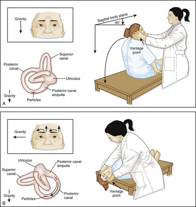
Box 96.2 Findings on the Dix-Hallpike Test in Patients with Benign Paroxysmal Positional Vertigo
Latency is approximately 3 to 10 seconds.
Significant subjective vertigo is present.
The intensity of vertigo escalates and then slowly resolves.
Vertigo and nystagmus last for approximately 5 to 30 seconds.
The nystagmus is upbeat (toward the forehead) and torsional toward the abnormal ear.
The nystagmus often reverses direction when the patient returns to the seated position.
Fatigability—the vertigo and nystagmus decrease and eventually subside with repeated positioning.

Stay updated, free articles. Join our Telegram channel

Full access? Get Clinical Tree

