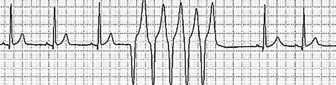
Fig. 17.1
Monomorphic ventricular tachycardia
Question
What approach should be taken in management of multiple recurrences of ventricular arrhythmias over a short period of time, i.e. VT storm?
Answer
Antiarrhythmic medications and correction of the arrhythmia trigger(s).
The patient was initially treated with intravenous amiodarone and external defibrillation for episodes of VT. A temporary transvenous pacemaker was inserted to prevent bradycardia in the setting of intermittent complete atrioventricular block. Electrolytes were repleted. Intravenous lidocaine was added for breakthrough episodes of VT. Benzodiazepines were administered to ameliorate anxiety. Echocardiography showed left ventricular systolic dysfunction, with an estimated ejection fraction of 30 %. Right heart catheterization revealed elevated intracardiac filling pressures and a cardiac index of 1.9 L/min/m2. An endomyocardial biopsy was negative for signs of inflammation or myocarditis. Left heart catheterization demonstrated severe native three vessel disease with an atretic left internal mammary artery (LIMA) graft to the left anterior descending (LAD) coronary artery, and patent vein grafts to obtuse marginal (OM2), diagonal branch (D1), and right coronary artery (RCA). A drug eluting stent was placed in mid LAD. There was no significant elevation in serum troponin levels. No further episodes of VT were observed, even after discontinuation of lidocaine. The patient received an implantable cardioverter defibrillator (ICD) and was discharged home on a long-acting beta blocker.
Three weeks later the patient presented with pre-syncope and palpitations. ICD interrogation revealed multiple episodes of VT resulting in defibrillator shocks. Intravenous amiodarone was initiated, but VT continued to recur. An intra-aortic balloon pump was placed with subsequent resolution of the VT. He then underwent electro-anatomic mapping followed by radiofrequency catheter ablation, after which he had no further VT.
Principles of Management
Diagnosis
Ventricular arrhythmias include a broad spectrum of rhythm disorders, which may have no clinical consequence or may result in sudden cardiac death. Patients at high risk for ventricular arrhythmias are those with ischemic heart disease, myocarditis, underlying structural heart disease, inherited or acquired channelopathies, drug intoxication, electrolyte derangement, and hyperthyroidism (Table 17.1). However, ventricular arrhythmias may occur in the absence of any of these predisposing factors. The diagnosis of ventricular arrhythmias may be suggested by the clinical presentation, but confirmation requires electrocardiographic recording.
Table 17.1
Approach to common etiologies of ventricular arrhythmias
Etiology | History | Evaluation | Treatment |
|---|---|---|---|
Myocardial ischemia | Chest pain, dyspnea | ECG, echocardiography, stress testing, coronary angiography | Coronary revascularization, beta blockers, intra-aortic balloon pump, ablation |
Channelopathies | Syncope, family history of sudden death | ECG, exercise testing, provocative pharmacological testing | Removal of QT-prolonging drugs, intravenous magnesium, potassium replacement, temporary pacing, sympathectomy, ICD |
Myocarditis | Flu-like symptoms, chest pain | ECG, echocardiography, cardiac MRI, endomyocardial biopsy | Amiodarone, beta blockers, ICD, steroids in selected cases where indicated for subtypes of myocarditis |
Electrolyte imbalance | Renal failure, dehydration | Serum electrolytes, ECG | Correction of electrolytes |
Cardiomyopathy | Symptoms of heart failure | Echocardiography, coronary angiography, cardiac MRI | Amiodarone, beta blockers, ICD |
Sarcoidosis | Lung involvement | Cardiac MRI, biopsy | Steroid therapy, immunomodulating agents, ICD |
Arrhythmogenic right ventricular cardiomyopathy | Palpitations, syncope, dyspnea, family history | ECG, cardiac MRI, genetic testing | Beta blockers, ICD, amiodarone |
Drug intoxication | Drug abuse, use of QT prolonging medications or digoxin | ECG, serum drug levels | Stop offending agent |
Hyperthyroidism | Anxiety, palpitations, heat intolerance | Thyroid hormone studies | Beta blockers, glucocorticoids, anti-thyroid medications |
Classification of VT
Ventricular arrhythmias are subdivided into non-sustained (premature ventricular contractions and non-sustained VT) and sustained (defined as VT lasting for more than 30 s or VF). VT may be monomorphic or polymorphic, with torsades de pointes representing a specific form of polymorphic VT occurring in the setting of a prolonged QT interval. Electrical storm is defined as 3 or more episodes of sustained VT, VF, or appropriate ICD therapies during a 24-h period. This condition has variably been referred to as “VT storm” or “VT cluster”. An important subgroup of electrical storm is incessant VT, which refers to repeated recurrence within 5 min of a technically successful therapy. Management of sustained ventricular arrhythmias is essential aspect of critical care medicine.
Monitoring and Testing
Patients presenting with a suspicion of ventricular arrhythmias should undergo continuous electrocardiographic monitoring in a setting in which appropriate care can be rapidly deployed. A 12-lead ECG should be obtained at baseline and assessed for evidence of ischemia, underlying structural heart disease, and the various channelopathies, such as long QT syndrome or Brugada syndrome (Fig. 17.2). Every attempt should be made to obtain a 12-lead ECG during the occurrence of a ventricular arrhythmia. Confirmation of the presence of a ventricular arrhythmia may require prolonged electrocardiographic monitoring (inpatient or outpatient) (Fig. 17.3), or invasive electrophysiologic testing. Once the diagnosis of a ventricular arrhythmia has been confirmed, further studies should be obtained as clinically indicated in order to elucidate the underlying cause. These may include: serum electrolyte testing, thyroid studies, screening for drugs of abuse, echocardiography, cardiac magnetic resonance imaging, coronary angiography, endomyocardial biopsy, exercise electrocardiography, and provocative pharmacological testing. A family medical history should be assessed for sudden death or known cardiomyopathies. Myocardial ischemia should be considered in all cases of polymorphic VT and ventricular fibrillation with coronary angiography being appropriate early in the evaluation in most cases.
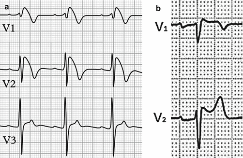
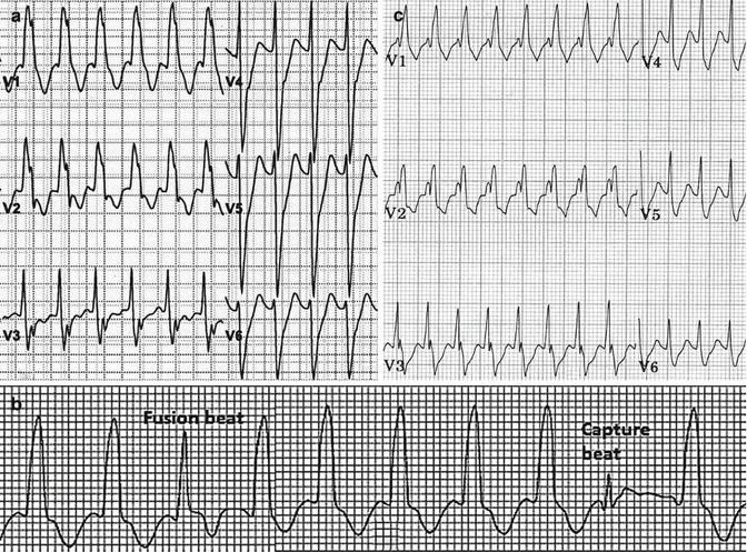
Fig. 17.2
Electrocardiographic tracings in Brugada syndrome. (a) Type 1; “coved type” ST-T segment configuration. (b) Type 2; “saddle back” pattern
Fig. 17.3
(a) Monomorphic ventricular tachycardia. Atrioventricular dissociation can be seen. (b) Ventricular tachycardia with fusion beat (hybrid complex of supraventricular and ventricular activation) followed by a capture beat. (c) Supraventricular tachycardia with aberrancy. Atrioventricular dissociation and fusion beats are absent. RS complexes in precordial leads are evident
General Measures
Non-sustained VT (Fig. 17.4) is encountered very commonly in the intensive care unit among patients with structural heart disease. In general, no specific therapy is required for asymptomatic patients other than electrolyte repletion and treatment of the underlying condition. For frequent or symptomatic non-sustained VT, however, treatment with beta blockers should be considered. Patients who are not otherwise candidates for an ICD for primary prevention of sudden death, for example, those with non-sustained VT, a history of MI, and moderate left ventricular systolic dysfunction (EF >35 to 40 %), may be considered for electrophysiology study (EPS) when stable in order to determine if an ICD is indicated.