Absent distal pulses
Expanding hematoma
Pulsatile bleeding
Palpable thrill
Audible bruit
In rare cases, with an expanding hematoma in an extremity, no further evaluation should take place, and the patient should be taken to the operating room to stop the bleeding, preferably by proximal control or direct exploration. If free pulsatile bleeding is obvious from the open fracture, tamponade is done as quickly as possible and a tourniquet should be considered. Recent experiences in the Iraq and Afghanistan conflicts show good results in these devastating events [4, 5].
20.2.2 Doppler Evaluation
In many occasions, Doppler evaluation is done for further evaluation of the vascular system. However, Doppler evaluation is only a valuable tool, if it is accompanied with a Doppler-guided pressure reading and an ankle-brachial index evaluation. The ankle-brachial index should be above 90 % to exclude vascular injury [6]. Picking up a Doppler-positive signal does not exclude a major vascular compromise, as in most times some signal is picked up from collaterals, which are however insignificant for the survival of the extremity. Only very experienced vascular surgeons or vascular technicians can evaluate the spectrum of the Doppler signal in such cases, although they mostly rely on a formal spectral analysis.
20.2.3 Angiography
Angiography is the gold standard in the evaluation of vascular injuries (Fig. 20.1) [7]. Depending on the urgency or complexity of the case, this can be done either in the angio suite, which is in many cases preferable because of the extensive and high quality radiological possibilities, or in the OR, most times with less sophisticated equipment. The OR environment, however, is favorable in patients with multisystem injuries or for instance in damage control situations [8]. A simple one-shot angiogram through a proximal arterial puncture gives most times a very adequate overview of the vascular system and the level of the problem.
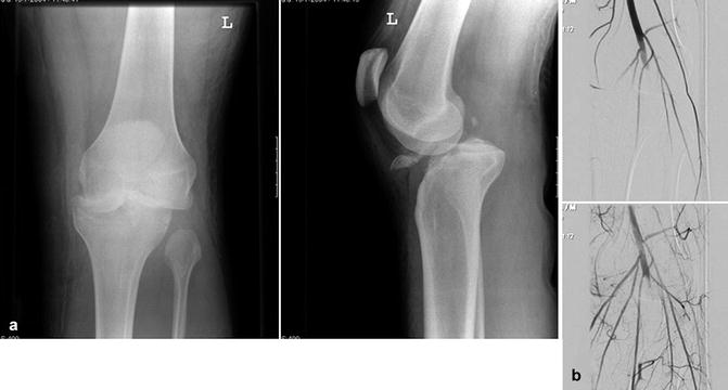

Fig. 20.1
(a) Patient with a knee dislocation. (b) Subsequent arteriography demonstrated a complete stop at the popliteal artery
Advantage of the arteriography is the possibility of angio embolization. In cases of severe arterial bleeding, e.g., in pelvic fractures, angio embolization can be an important adjunct in the treatment of these severely injured patients, after initial mechanical stabilization and packing. Intraluminal manipulation, when performing an angiogram, gives also the possibility of using intraluminal stents. These stents can be used for bridging defects, occluded trajectories and coverage of traumatic pseudoaneurysms [9]. The use of large amounts of intravenous contrast has the disadvantage of a chance of contrast nephropathy and allergic reaction. In emergency cases also, the chance of local vessel injury is relevant [10].
20.2.4 CT Angiogram
As in the current practice, CT is very often used for the evaluation of the trauma patient. CT angiography is an option for further evaluation of the vascular status of the trauma patient. A specific protocol and timing should be used for an optimal result. This modality is less invasive compared to the classic angiogram; however, contrast related problems can occur as well with this technique. It has largely replaced the invasive angiography for initial diagnostics in the trauma setting as it is readily available [11].
20.2.5 Digital Subtraction Angiography
Intravenous digital subtraction angiography (DSA) can be used in selected cases; however, it results in inferior image quality and requires a trip to the radiology department. In children, however, this can be an option, as the vascular system is less easy to catheterize (Fig. 20.2). The relative high dose of contrast that is given is the disadvantage of this technique.
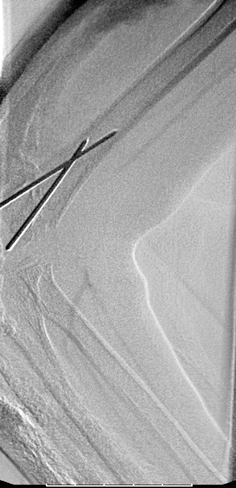

Fig. 20.2
Digital intravenous subtraction angiography in a child with a supracondylar humeral fracture. Disruption of the brachial artery in the area of the fracture fixed with two K-wires
20.2.6 MR Angiography
Increasing popular in vascular surgery is the use of the MR angiography. Because of the very specific circumstances where the multiply injured patient is many times on the ventilator, this modality is, infrequently used in the early evaluation of the trauma patient [7].
20.3 Treatment
20.3.1 General Tactics
20.3.1.1 Logistics
In the last years, major changes in the logistics for the optimal care of the trauma patient have been imminent. Not only the further advancement of the CT, even into the emergency room and even used in hemodynamically unstable patient, but also the further development of the endovascular techniques in vascular surgery led to the development of hybrid operation suites in which classic operative and endovascular techniques could be used in unison. This development is of major importance in patients with multisystem injuries with skeletal, visceral and vascular lesions. In such a suite, all modalities can be used for optimal care of the trauma patient without the necessity to transport the patient, with all its dangers and intricacies. For instance, patients with major pelvic injuries after initial mechanical stabilization and packing can undergo catheter-guided embolization in the same instance and place. Moreover, patients with severe hepatic injuries can undergo after initial packing embolization of the remaining arterial intrahepatic lesions, without dangerous transport to the angio suite.
20.3.2 Severe Torso Bleeding
Non-compressible torso bleeding still remains a major cause of death in the trauma patient. Morrison and coworkers [12] reported the use of the resuscitative endovascular balloon occlusion of the aorta in severe torso trauma with impressive experimental results, reducing mortality to 25 % with continuous balloon occlusion. In a clinical series of both blunt and penetrating injury, Brenner and coworkers [13] reached hemorrhage control by aortic balloon occlusion through percutaneous or direct cut-down of the femoral artery. There was no hemorrhage-related death in this small series, with descending aortic and infrarenal aortic lesions. New aortic balloon systems can be used fluoroscopy free; therefore, it is suitable for use in the mergence room or trauma bay.
20.3.3 Extremity Bleeding
Several tactics can be chosen after the diagnosis is obvious. For an overview, see the treatment algorithm. In severe open wounds with heavy bleeding, tamponade is the treatment of choice. This can be manually done (Fig. 20.8). Recent experiences in the Iraq and Afghanistan conflicts showed a renewed interest and good results of the application of tourniquets, as mentioned above.
After the prehospital and initial resuscitation phase, gaining proximal control is of utmost importance. Thereafter, revascularization is done as soon as possible. In case of complex combined vascular and musculoskeletal injuries, regaining perfusion of the distal part of the extremity is very important. Nevertheless, it should not compromise the possibilities for orthopedic intervention neither should the orthopedic intervention make an adequate vascular procedure impossible. Although 6 h of ischemia time is tolerated in an injured leg, as little ischemia time as possible should be allowed. The longer the ischemia time in an injured leg, the higher the coagulation disposition will be. An adequate option is to use a shunt (Fig. 20.3) to bridge the time to definitive care with a well-perfused distal part of the extremity.
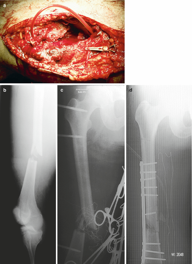

Fig. 20.3
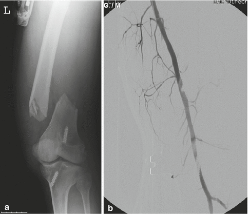
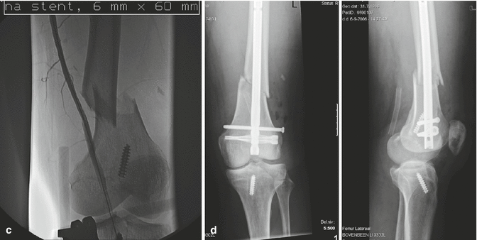
(a) Shunt in situ in the superficial femoral artery in a patient with a femoral fracture and severe head injury. After hemorrhage control, restoration of flow by (b) a shunt, (c) temporary external fixation, and (d) ultimate plate fixation of the femur
From the orthopedic standpoint, temporary stabilization of the fracture with an external fixator is a good option. It shortens the time to vascular reconstruction as well as reperfusion and leaves the opportunity for extensive reconstruction after the vascular continuity is restored. Care should be taken to restore adequate length, so the definitive reconstruction of the bone can be done without major shortening or lengthening of the extremity, as this can compromise the vascular conduit later on.
For repair, mostly an interposition vein graft is used [14], because of the immunological properties (Fig. 20.8). A PTFE conduit can be used; however in case of open fractures and contaminated wounds, this is the less preferable option. Direct repair can be used in selected cases; however in order to prevent a relevant stenosis after direct repair, a vein patch is often used instead [15].
In case of an incomplete occlusion of an artery, most times based on a stretching mechanism, with an intimal tear as the result, several options are available [16]. Antiplatelet therapy has been advised in such cases, e.g., in case of carotid artery lesions after cervical fractures [17]. Other authors advocate the use of a wall stent, placed through radiological intervention (Fig. 20.4). Stents have been used for a variety of vascular problems like aneurysms, dissections and hematoma [18].

