UPPER EXTREMITY FRACTURES: ORTHOPEDIC MANAGEMENT

CHAPTER 67 UPPER EXTREMITY FRACTURES: ORTHOPEDIC MANAGEMENT


Fractures and dislocations of the upper extremity can vary from benign, requiring minimal intervention, to life and limb threatening. The treatment plan is based on the injury pattern including location, associated neurologic or vascular injury, status of the soft tissues, mechanism, and other associated injuries. In this chapter, several key issues in the decision-making process are discussed, followed by description and treatment of specific injuries.


OPEN FRACTURES


The associated soft tissue injuries add an element of urgency to the treatment of fractures. A fracture is classified as “open” if the fracture or fracture hematoma communicates with the air via a wound in the soft tissues. This can be caused from the bone protruding through the skin, “inside out,” or if there is a penetrating mechanism causing an injury from the “outside in.” Regardless, the implication is that environmental contamination can increase the incidence of infection and fracture healing complications. If there is a wound in the same limb segment as the fracture, it should be considered open until proven otherwise. A classification system for open fractures appears in Table 1. Infection rates are reported at 0%–2% for Type I, 2%–7% for Type II, and 10%–25% for Type III overall. Rates for type III are subclassified as follows: IIIA, 7%; IIIB, 10%–50%; and IIIC, 25%–50%.


Table 1 Classification of Open Fractures


Treatment of these fractures is therefore aimed at irrigation and debridement in the operating room within 8 hours. Preliminary or definitive stabilization of the fracture and appropriate antibiotic therapy should follow. It is apparent that short-course, high-dose antibiotic therapy is appropriate for open fractures and need not be continued over the course of the fracture healing process. Recommendations follow:


Trauma to the arm involving a severe crush component such as that from a conveyer belt or injuries with prolonged vascular compromised are at risk for development of compartment syndrome. Prophylactic fasciotomy may be necessary.


DISLOCATIONS


This is another situation that requires more urgent assessment, diagnosis, and treatment. By definition a dislocation is present when the joint is disrupted such that the articular surfaces are no longer in contact. The diagnosis of a joint dislocation is often made from the history and physical examination. The limb will usually be held in a fixed position characteristic of the dislocation direction. A posterior hip dislocation, for example, is seen with the limb in flexion, adduction, and internal rotation. Loss of the normal contour of the joint can be seen as evidenced by a “sulcus” sign in an anterior shoulder dislocation.


Radiographic evaluation is essential in the management of these injuries because associated fractures will otherwise go unrecognized and can have significant effects on the prognosis if not taken into account before reduction.


Neurovascular compromise is the reason for emergent reduction of the joint. Sciatic nerve injury has been reported to occur in 8%–19% of hip dislocations. Osteonecrosis is a known complication of hip dislocations as well, occurring in up to 17% of these injuries. This is due to the interruption of capsular blood supply from the increased tension caused by the dislocation. Other associated neurologic injuries can be seen in Table 2.


Table 2 Neurologic Injuries Associated with Upper Extremity Fractures


Joint Common Neurologic Injury Deficit
Shoulder Axillary nerve Sensory deficit in deltoid region, weakness of deltoid and teres minor
Elbow Posterior interosseous nerve Weakness of wrist dorsiflexion
Knee Peroneal nerve Weakness of ankle and great toe dorsiflexion
Hip Sciatic nerve More frequently common peroneal portion giving dorsiflexion weakness

Dislocations and fractures with neurologic or vascular compromise should therefore be reduced as quickly as possible in order to reduce potential irreversible injury to the affected structures. Following reduction a repeat examination is warranted to see if there has been a change in the neurovascular status of the limb.


GUNSHOT WOUNDS


Special attention is deserved here due to the need to make an important distinction for the treatment of these injuries. It is important to determine if the wound was inflicted by a “low-” or “high-velocity” weapon. The exact distinction is somewhat cloudy, but according to the Wound Ballistics Manual of the Office of the Surgeon General, muzzle velocity greater than 2500 ft/sec constitutes “high velocity.” This is important because the kinetic energy of the bullet varies directly with the square of its velocity and only linearly with its mass.


Many low-velocity gunshot injuries can be treated to completion with closed methods, such as functional bracing or casting. If one chooses open management, it should be anticipated that the extent of the fracture is often more extensive than can be appreciated on plain radiographs. This should be taken into account in the preoperative planning.


There may also be associated neurologic deficits due to the “blast effect” of the initial injury that do not warrant immediate exploration, as many will often resolve. If persistent neurologic deficit occurs, an electromyograph (EMG) may be indicated at 3–6 weeks to assess the nerve for any evidence of fibrillation potential.


Gunshots to the forearm require special attention even if there is no associated fracture due to an increased risk for development of forearm compartment syndrome. During the past 4 years, we have treated five patients who developed forearm compartment syndrome from penetrating injuries. All were found to have arterial lacerations. Patients should be monitored for at least 8–12 hours for clinical evidence of increased pressure within the forearm, which include marked pain on passive digital extension, tense or swollen forearm, and reduced hand sensibility or paresthesias. Intracompartmental pressure measurements may be helpful but the diagnosis is made on clinical grounds.


Once the diagnosis is established, an emergent forearm fasciotomy is required to prevent Volkmann’s contracture.


INJURIES TO SHOULDER GIRDLE AND HUMERUS


Scapula Fractures


Fractures of the scapula are relatively uncommon, accounting for 3% of all shoulder girdle injuries. These generally occur as the result of high-energy trauma explaining the frequent association with other, often life-threatening, injuries that may be of greater significance than the fracture itself. It is not uncommon for scapular fractures to be overlooked in polytrauma patients and often noticed incidentally on a chest radiograph or CAT scan.


A classification system was developed by Ada and Miller (Table 3), which divided fractures into those involving the acromion, spine and coracoid, type 1, glenoid neck, type 2, intra-articular glenoid fractures, type 3, and isolated scapular body fractures, type 4.


Table 3 Scapular Fracture Classification System


Fracture Type Anatomic Description/Location
1A Fracture through acromion
1B Fracture line through base of acromion or scapular spine
1C Fracture through coracoid process
2A Vertical glenoid neck fracture, lateral to base of acromion
2B Vertical glenoid neck fracture that extends up through scapular spine and supraspinatus fossa
2C Fracture line starts laterally at glenoid neck and propagates in transverse fashion through body exiting medially
3 Intra-articular glenoid fracture
4 Scapular body only

Data from Ada JR, Miller ME: Scapular fractures: analysis of 113 cases. Clin Orthop 269:174–180, 1991.


Associated injuries are quite common due to the high-energy mechanisms in which they usually occur. In one study of 148 fractures in 116 scapulae, 96% had associated injuries with upper thoracic rib fractures being the most common. Pulmonary injuries were also common with an overall incidence of 37%, of which 29% were hemopneumothorax and 8% pulmonary contusion. Head injuries were observed in 34%, ipsilateral clavicle fractures were seen in 25%, and 12% of patients had cervical spine injuries, of which 4% had permanent cord injuries.


Management of these fractures is often nonsurgical as poor healing is an infrequent complication due to the rich blood supply from the investing rotator cuff musculature. Nonoperative management consists of admission for a period of 24 hours to assess pulmonary and cardiac status. A brief period of sling immobilization is initiated for comfort, followed by passive range of motion. Most fractures are united by 6 weeks such that active mobilization and strengthening can ensue safely. Maximal functional recovery can take 6–12 months.


Operative indications include intra-articular glenoid fractures with 5 mm of displacement, coracoid fractures with intra-articular extension and more than 5 mm of step-off, glenoid rim fractures with persistent or recurrent glenohumeral instability.


The injury pattern described as the “floating shoulder” deserves attention. This refers to a “double disruption” of the superior shoulder suspensory complex. This complex consists of a bone and soft tissue ring formed by the glenoid, coracoid process, coracoclavicular ligaments, distal clavicle, acromioclavicular joint, and acromion process. Isolated disruption of one of these components is generally tolerated well; however, when two or more structures are damaged, it is thought to produce an unstable situation. This is commonly seen with ipsilateral clavicle and glenoid neck fractures. The treatment of such injuries is somewhat controversial in that good results have been reported with fixation of both the glenoid and clavicle, clavicle alone, and more recently, nonoperative management. At this time, reasonable indications for operative intervention would include glenoid neck fractures with more than 3 cm of medial displacement in combination with injury to another structure. Each patient, however, must be evaluated individually.


Scapulothoracic Dissociation


Scapulothoracic dissociation is an infrequent injury that can be thought of as an internal forequarter amputation, which is almost always seen in conjunction with severe injuries to the brachial plexus and subclavian vessels. It is the result of a massive traction injury causing significant lateral displacement of the scapula relative to its thoracic articulation. According to the paper by Althausen et al., 88% have associated vascular lesions and 94% presented with severe neurologic injuries. A flail extremity resulted in approximately 52%, early amputation in 21%, and death in 10% of patients.


Diagnosis is made clinically by the presence of massive swelling, weakness, pain, tenderness, and absent or diminished pulses. It is crucial not to attribute pulselessness to a more distal injury when a more proximal, life threatening injury may be the underlying cause. Radiographically an AP film of the chest with the cassette oriented transversely may reveal lateral displacement of the scapula. Measurement from the medial border of the scapula to the spinous processes should alert the physician to the possibility of a scapulothoracic dissociation with a distance of more than 1 cm when compared to the opposite side. This may or may not be seen in conjunction with an acromioclavicular separation or clavicle fracture.


Management is controversial but arterial injuries may warrant immediate exploration and repair. As many as 10% of patients who survive the initial injury may die from exsanguination. Brachial plexus exploration may be carried out at the same time. Bony stabilization may be necessary to protect vascular repairs but the role of internal fixation is otherwise less clear.


Glenohumeral Dislocation


Due to its lack of bony constraint, the shoulder is the most commonly dislocated joint in the body. It is not a true ball and socket joint, but more like a golf ball resting on a golf tee. The static restraints to dislocation are composed of the glenoid labrum, capsule, and glenohumeral ligaments, while the rotator cuff musculature provides additional dynamic stability.


Anterior dislocations are by far the most common type. These are usually the result of an eccentric load applied to the arm while in an outstretched position as would be seen in a volleyball player while spiking the ball. Posterior dislocations occur with a posteriorly directed force on an adducted, flexed arm, and also have been noted to occur in patients who are seizing.


An anteriorly dislocated shoulder will present with the arm at the side or in slight abduction and external rotation. Normal loss of the shoulder contour may be seen with a prominent “sulcus” sign. Adduction and internal rotation are usually limited. A patient with a posterior dislocation will hold the arm in an adducted, internally rotated position.


Evaluation should include a thorough neurologic examination prior to any attempted reduction. Neurologic involvement is not infrequent with the axillary nerve being most commonly affected. Vascular status should likewise be documented, as vascular injuries can occur, although less commonly. Radiographic evaluation should also be completed prior to commencing treatment. A standard trauma series as described earlier is extremely helpful in delineating any associated glenoid rim or humeral head impaction fractures.


Reduction is most easily carried out with some form of sedation and or injection of lidocaine into the joint capsule. Gentle traction–counter traction will reduce most dislocations. Irreducible dislocations and fracture dislocations are best managed in the operating room with general anesthesia.


Postreduction, a period of immobilization in a sling from 10 days to 2 weeks is recommended followed by a supervised physical therapy program. In patients younger than 20 years, recurrence rates up to 90% have been reported most likely due to the violent nature of the dislocation and the very commonly associated anterior-inferior labral tear or “Bankart” lesion that occurs. In contrast, patients aged over 40 years commonly have associated rotator cuff tears. A high index of suspicion should be present at follow-up examination of these patients in the early recovery period.


Proximal Humerus Fractures


Proximal humerus fractures are common injuries, especially with our aging population. The majority of these injuries are minimally displaced or nondisplaced and can be treated conservatively. Factors to take into consideration in the treatment plan are age of the patient, hand dominance, bone quality, fracture type, and fracture displacement. Associated injuries in the multitrauma patient are also important in the decision-making process.


Assessment should consist of a thorough neurovascular examination along with a radiographic trauma series. The presence of an expanding axillary mass and absent distal pulses is concerning for a vascular injury. Nerve injuries occur in as many as one-third of patients and are more common with increasing age. In one study the incidence of nerve injury with proximal humeral fracture-dislocations was greater than 50% after age 50. If present, these injuries may take 9 months or more to recover neurologic function, which is often incomplete.


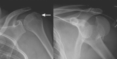
These fractures are classified according to the description by Neer. He described six variations of displaced proximal humerus fractures, and defined displacement as greater than 1 cm or 45 degrees of angulation. The anatomic “parts” consist of the anatomic neck, surgical neck, and greater and lesser tuberosities. Despite poor interobserver reliability, this is the most frequently used classification system. Figure 2 depicts various fracture patterns that are both considered “two-part fractures.” The portion of the humerus that is displaced has clinical relevance due to the varying blood supply of the proximal humerus. Displacement of the anatomic neck for example, has a high chance of disrupting the blood supply and adversely affecting outcome, regardless of the treatment chosen.


< div class='tao-gold-member'>

Stay updated, free articles. Join our Telegram channel

Jul 7, 2016 | Posted by in CRITICAL CARE | Comments Off on UPPER EXTREMITY FRACTURES: ORTHOPEDIC MANAGEMENT

Full access? Get Clinical Tree

Get Clinical Tree app for offline access