CHAPTER 47 GASTRIC INJURIES
The stomach is a relatively thick-walled, well-vascularized organ that is variably positioned in the peritoneal cavity. Although partially protected by the lower rib cage, its size and location put the stomach at risk for injury, particularly with injury from penetrating trauma to the abdomen or lower chest.
INCIDENCE
Gastric injuries usually result from penetrating trauma and occur in approximately 20% of gunshot wounds and 10% of stab wounds.1–3 Blunt gastric trauma is much less common. The American Association for the Surgery of Trauma (East) multi-institutional study on hollow viscus injury reported that the prevalence of blunt gastric rupture was 0.06% in patients undergoing evaluation for blunt abdominal trauma and 2.1% of all patients found to have hollow viscus injury.4
MECHANISM OF INJURY
The stomach is at risk for injury after stab wounds to the left thoracoabdominal region of the body. A single perforation occurs in over 50% of these cases.2 However, injury to adjacent organs is common. Gunshot wounds result in two or more gastric wounds in 90% of cases.2 Although often associated with some surrounding tissue damage to the stomach, this is usually only significant with high-velocity missiles. Shotgun wounds at close range (<15 feet) are often associated with massive destruction of the abdominal wall, stomach, and other intra-abdominal organs.
Blunt injury to the stomach is most often the result of motor vehicle crashes, or motor vehicle–pedestrian trauma.5–7 Less common causes include falls, assaults, and improperly performed cardiopulmonary resuscitation. Blunt gastric injuries include linear lacerations and complete gastric rupture. The postulated mechanisms for blunt gastric injury include sudden increases in intraluminal pressure resulting in a balloon-bursting type of phenomenon of a full stomach, compression against the spine (seat-belt injury), or a deceleration injury with shearing forces resulting in a laceration of the anterior stomach wall.
DIAGNOSIS
Patients with stab wounds and hypotension, peritonitis, or both should undergo laparotomy immediately. Asymptomatic patients without central nervous system injury (brain or spinal cord injury) or drug or alcohol involvement may be observed with repeated physical exams. In other patients, local wound exploration, diagnostic peritoneal lavage (DPL), or laparoscopy are alternatives. Laparoscopy is most helpful with thoracoabdominal stab wounds in identifying associated injuries to the diaphragm.8 Focused assessment with sonography for trauma (FAST) may not identify the small amount of fluid initially associated with hollow viscus injury, and thus may be misleading with isolated gastric injuries.
Early operation is indicated for symptomatic gunshot wounds to the abdomen. Occasionally, a tangential gunshot wound in a stable patient may be observed, or such a wound may be found after the patient undergoes either DPL or laparoscopy. Abdominal CT is also helpful in this situation. In patients suspected to have either blunt or penetrating gastric injury, the placement of a nasogastric tube is helpful. Not only does proper placement of a nasogastric tube minimize the risk of aspiration, but a bloody aspirate when present is highly suspicious for a gastric injury. In patients with blunt gastric injury and no obvious peritoneal signs, a supine film of the abdomen discloses free air in less than 50% of cases. In this situation, abdominal CT is more sensitive in identifying free air.
SURGICAL MANAGEMENT
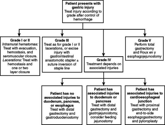
Gastric injuries thus identified are treated according to their severity (Table 1, Figure 1). Most intramural hematomas (grades I and II) are treated by careful evacuation, hemostasis, and closure with seromuscular sutures made of nonabsorbable material. Small grade I and II perforations can be closed in one or two layers. Because of the vascularity of the stomach, I prefer a two-layer closure after hemostasis is achieved.
Table 1 AAST Organ Injury Scale for Stomach
| AAST Grade | Characteristics of Injury |
|---|---|
| I | Intramural hematoma <3 cm, partial thickness laceration |
| II | Intramural hematoma >3 cm; small (<3 cm) laceration |
| III | Large (>3 cm) laceration |
| IV | Large laceration involving vessels of greater or lesser curvature |
| V | Extensive (>50%) rupture; stomach devascularization |
AAST, American Association for the Surgery of Trauma.
Modified from American Association for the Surgery of Trauma (AAST).
< div class='tao-gold-member'>
Related posts:


Stay updated, free articles. Join our Telegram channel

Full access? Get Clinical Tree


