CHAPTER 24 TRAUMATIC BRAIN INJURY: PATHOPHYSIOLOGY, CLINICAL DIAGNOSIS, AND PREHOSPITAL AND EMERGENCY CENTER CARE
Death. Long-lasting or even permanent loss of function. Those are the burdens borne by many traumatic brain injury (TBI) patients and their families. Even some patients who initially appeared to have injuries that were mild according to clinical or radiographic criteria can suffer permanent injury.
Emergency craniotomies and insertion of intracranial monitors are the most high-profile aspects of management of TBI patients. However, the vulnerability of the injured brain to even mild and transient metabolic derangements underscores the major impact that systemic parameters can have on influencing outcome from TBI. Thus, non-neurosurgeons can influence management in ways that are just as important, and in some cases perhaps more so, than the interventions performed by neurosurgeons.
This chapter will discuss a few principles of the underlying pathophysiology, initial assessment, and prehospital and emergency center management of TBI patients. The following chapter addresses topics relevant to the acute hospital admission. This discussion is weighted toward patients with severe TBI, but many of the basic principles apply to patients with mild or moderate TBI as well.
INCIDENCE
It is frequently stated that in multiply injured patients, the head is the most commonly injured part of the body. Outcome from polytrauma is more dependent on the extent of brain injury than on injury to other organ systems. Perhaps a third of the entire cost of trauma, including medical and rehabilitative care, lost income to the patient, and lost productivity to society, is attributable to brain injury.
According to data reported by the Centers for Disease Control and Prevention in the 1990s, 1.5 million Americans sustain a TBI every year. Of this number, hospitalization and ultimate survival occur in approximately 230,000 patients, but 50,000 will die. Long-term disability will occur in 80,000-90,000 patients annually. It has been estimated that more than 5 million men, women, and children in the United States are living with a permanent TBI-related disability.1
The TBI death rate in the United States is approximately 20 per 100,000 population. In all age groups, the mortality rate is higher in males than in females. The incidence of TBI-related mortality peaks in the late teens and early twenties, subsequently decreasing during the next few decades until taking off exponentially at about retirement age.
MECHANISM OF INJURY
Although an epidural, subdural, or intraparenchymal hematoma may have a dramatic appearance on a computed tomography (CT) scan, the clinician must remember that these lesions are distinct from the cerebral parenchymal injury that is the true cause of long-term neurologic deficits. TBI is best thought of as a diffuse disturbance of cerebral function, not as a blood clot or contusion. This diffuse disturbance may occur in parallel with, but may also be independent of, those processes that lead to the development of traumatic mass lesions.
Subdural Hematoma
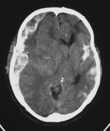
Classically, a subdural hematoma (SDH) (Figure 1) has been said to develop after tearing of a bridging vein, that is, a vein passing directly from the cortex to the overlying dura. The mechanical forces of the trauma can cause tearing of these veins. More recent evidence indicates that at least some of these hematomas actually form from splitting of inner and outer layers of the dura, that is, they may actually be “intradural hematomas.” Finally, some SDHs are caused by direct bleeding into the subdural space from parenchymal contusions or hematomas or from injured cortical arteries or veins.
Epidural Hematoma
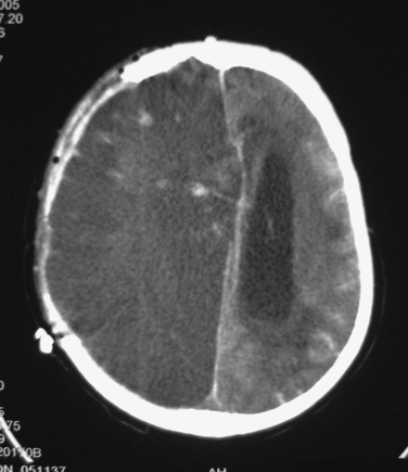
Epidural hematomas (EDHs) classically arise after a blow to the side of the head results in a fracture of the thin temporal bone immediately overlying the middle meningeal artery. The patient may briefly lose consciousness after the initial impact, but he or she quickly awakens; thus, the brain injury was mild. Unfortunately, the fracturing of the skull lacerated the middle meningeal artery. Continued bleeding from this source produces an enlarging EDH, the presence of which may be signaled by such symptoms as severe and worsening headache, vomiting, and decreasing level of consciousness. The period between awakening from the initial concussion and subsequent lapsing into a coma has historically been described as a “lucid interval.” Importantly, loss of consciousness does not always occur after the skull is fractured, and many patients with large EDHs are awake until they begin to lapse into a terminal coma. It must also be mentioned that many EDHs are not associated with meningeal arterial bleeding. In these cases, perhaps the source of the hematoma is oozing from the overlying edges of fractured bone.
Referring again to the classic scenarios, patients with EDHs are said to fare better than patients with similarly sized SDHs. Why should this be so? The answer is that a “pure” EDH essentially represents a skull fracture with no direct parenchymal injury to the brain. On the other hand, the rotational forces that are said to be an important cause of SDHs via tearing of bridging veins may also cause widespread axonal injury, as discussed later. Thus, SDH is said to be associated with a greater burden of parenchymal injury, which explains the worse outcomes. Of course, this explanation refers only to the extreme ends of the spectrum of the pathophysiology of EDHs and SDHs. Many patients with EDHs will do poorly, while SDH patients often recover well from their injuries. Nevertheless, this explanation is a useful way to conceptualize the interactions between mass lesions and diffuse injury.
Subarachnoid Hemorrhage
The most common post-traumatic intracerebral hemorrhage is not a mass lesion, but rather diffuse subarachnoid hemorrhage (SAH) (see Figure 1). Several retrospective series report that SAH after TBI is independently associated with worse outcomes, but the mechanism that might explain such an association is unclear. In the acute setting, SAH does not seem to have much effect on patient management, which is driven instead by more immediately pressing concerns.
Parenchymal Lesions
Contusions occur commonly after TBI, especially at the base of the frontal lobes and at the anterior edges of the temporal lobes. The brain in these regions is said to continue moving over or into the skull base after the head suddenly stops moving after a violent blow or rapid rotational movement. Fortunately, most such contusions remain small and surgically insignificant. Emergency surgery may be required, however, for larger lesions or for smaller ones that subsequently enlarge.
Unlike contusions, in which extravasated blood mixes freely with brain tissue, parenchymal hematomas consist of solid blood clots within the brain itself. They occur less commonly than contusions. They tend to be more variable in their distribution.
Ischemia
Diffuse injury may be of several types. Ischemia is a common form of diffuse injury. In some cases, mass effect from a traumatic hematoma may cause elevated intracranial pressure (ICP) and local compression of underlying tissue that can lead, respectively, to global and local reduction of cerebral blood flow (CBF). However, post-traumatic cerebral ischemia may also occur when no mass lesion is present, especially very early after injury. Although the CT scans in these cases may be relatively unimpressive, these patients may be quite vulnerable to the effects of such secondary insults as hypotension or hypoxia. Over the subsequent hours and days, CBF usually increases, but the damage from early ischemia may have already been done long before the increase in CBF (Figure 2). Some centers have used xenon CT or perfusion CT to measure CBF and identify ischemia immediately after injury.
Diffuse Axonal Injury
Another common type of diffuse injury is diffuse axonal injury (DAI). Although the axonal disconnection that characterizes DAI is commonly thought to occur at the time of injury, such immediate loss of axonal continuity probably occurs only when an injury produces severe cerebral parenchymal disruption. Instead, it appears more likely that the rotational and mechanical forces that are operant during the traumatic event produce a focal impairment of axoplasmic flow, which, in turn, culminates in axonal disconnection several hours after injury.2 This slight delay creates hope that a therapeutic window may exist for the administration of a yet-to-be-developed treatment that would prevent loss of axonal integrity and function.
Cellular and Molecular Factors
At the cellular level, the abnormalities caused by TBI are numerous and complex. Release of glutamate and other excitatory neurotransmitters may lead to excessive neuronal depolarization and intracellular calcium influx, with activation of proteases and other processes that lead to cell death. Inadequate blood flow can cause a conversion from aerobic to anaerobic metabolism. The lactic acid that is produced lowers local tissue pH, and the consequent acidosis contributes to tissue injury and death. Trauma-induced apoptosis may promote further cell death. These and other biochemical and cellular processes take place against the backdrop of an individual patient’s genetic makeup; the presence of specific alleles for various genes may make an individual more or less susceptible to the damaging effects of various pathophysiologic processes.
CLINICAL DIAGNOSIS
Clinical Examination
Ideally, the severity of TBI is determined and classified according to a patient’s neurologic examination. The size and appearance of a mass lesion as seen on imaging studies are not as important as the effect that the lesion may be having on a patient’s neurologic function and level of alertness.
The single most important question in the evaluation of a potentially head-injured patient is whether he or she obeys simple one-step commands. A simple definition of coma is that a person will not do such things as hold up two fingers or stick out the tongue when asked to do so. Failure to obey commands is widely used as an indicator of the presence of severe TBI. Other simple but important observations are the type of movement exhibited by the patient (localization of noxious stimuli, withdrawal, flexion, extension, etc.); whether the right and left sides are symmetrical; the type of speech; the presence or absence of eye opening; and pupillary size, reactivity to light, and bilateral symmetry.
Because the nerve fibers that mediate pupillary constriction lie on the surface of the third cranial nerve, compression of this nerve by herniating brain tissue that is being displaced by a large mass lesion may cause inactivation of the pupilloconstricting fibers. The resulting pupil appears large and unable to constrict in response to bright light. This physical finding in a comatose TBI patient suggests that an immediate CT scan is needed to identify a large acute hematoma. However, fixed and dilated pupils may also be caused by brainstem ischemia or by direct ocular trauma.
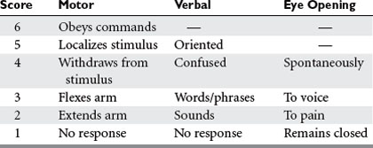
Many scales have been developed for the assessment of consciousness or neurologic status after injury, but by far the most widely used is the Glasgow Coma Scale (GCS)3 (Table 1). In conjunction with such information as the status of pupillary reactivity and the tempo or rate of change of a patient’s neurologic condition, the GCS is an extremely useful tool for assessing a patient’s baseline condition and subsequent progress.

Stay updated, free articles. Join our Telegram channel

Full access? Get Clinical Tree

