CHAPTER 21 THE USE OF COMPUTED TOMOGRAPHY IN INITIAL TRAUMA EVALUATION
In the 1970s, computed axial tomography (CT) revolutionized the evaluation and management of brain injury. Since then, advances in technology leading to increased speed and resolution have similarly changed the assessment of other injuries. Following the primary and secondary survey and initial plain radiographic evaluation, CT is often essential in the work-up of hemodynamically stable victims of blunt trauma. With the introduction of multidetector arrays, CT now may replace invasive catheter angiography and/or magnetic resonance imaging in certain circumstances.
COMPUTED TOMOGRAPHY OF HEAD/BRAIN (CRANIUM)
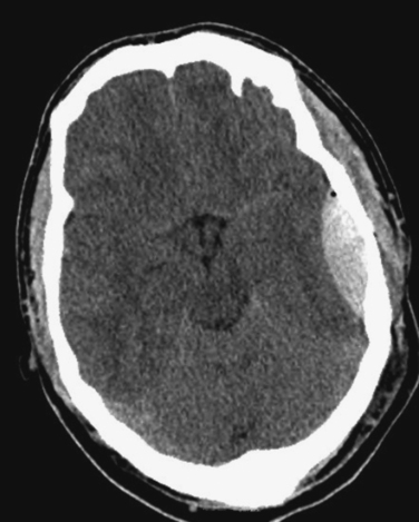
The study is performed without contrast. Acute hemorrhage appears white on the scan (Figure 1) except in very anemic patients, where it may appear isodense with brain. Active intracranial bleeding may also appear as gray swirls within the white clot, indicating hyperacute hemorrhage.

Figure 1 Computed tomography scan demonstrating a classic lenticular-shaped, acute epidural hematoma.
Computed tomography is very sensitive for intracranial hemorrhage, edema, and mass effect. As CT technology has advanced, detection of smaller lesions has become possible, leading to controversy regarding the significance of these lesions, and the indications for CT in mild head injury.1 CT of the brain may be normal and not correlate well with the clinical picture in patients with diffuse axonal injury.
COMPUTED TOMOGRAPHY OF FACE AND ORBITS
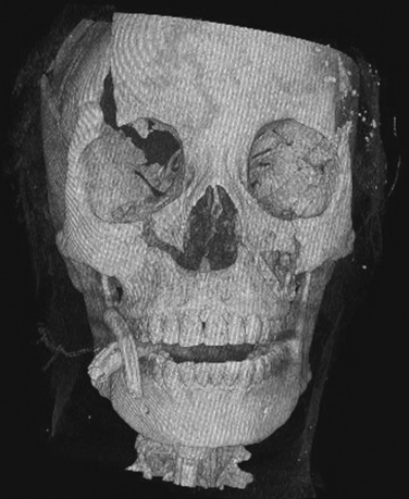
Axial images are obtained using 3-mm slices from below the mandible to just above the frontal sinuses. The mandible may be excluded if no suspicion of injury is present. In a spiral CT scanner, coronal views can be reconstructed from the initial data. With multidetector CT scanners, 3D reconstructions (Figure 2) can be generated that are of value to the maxillofacial surgeon in planning reconstruction, and particularly in complex fractures.
< div class='tao-gold-member'>
Stay updated, free articles. Join our Telegram channel

Full access? Get Clinical Tree


