Introduction
Vulvoscopy, the colposcopic examination of the vulva, is an essential part of an examination of a woman who presents with dyspareunia. A colposcope is a low-power microscope with an integrated light source that was initially developed to examine dysplastic and neoplastic lesions of the vagina and cervix. However, in the 1970s it was reported that the colposcope could also be used to improve visualization of the structures of the vulva. It was observed that colposcopy of the vulva (i.e., vulvoscopy) gave the examiner enhanced visualization of the labia majora, labia minora, interlabial sulci, vestibule, glans clitoris, prepuce, frenulum, hymen, minor vestibular glands, perineum, and anus.
Vulvoscopy enhances the ability to detect subtle colors changes associated with inflammatory or neo-plastic diseases of the vulva. Increased redness can be visualized when there are stromal changes due to inflammation, an immune response, or from neovascularization that occurs in association with neoplasia. In addition, there can be decreased vascularization, or fibrotic changes in the stroma, both of which appear pallid when viewed with a vulvoscope. Lastly, increased keratinization (lichenification) will appear as dense white areas of vulvar tissue.
Vulvoscopy can also help reveal scarring and architectural changes of the vulva. Chronic inflammatory disorders of the vulva, such as lichen sclerosus and lichen planus, frequently cause structural changes such as resorption of the labia minora, vulvar granuloma fissuratorum (chronic tearing of the posterior fourchette), and phimosis of the clitoris. These changes, which can be subtle, are best visualized with a vulvoscope.
In addition, inflammatory or dysplastic disorders of the vulva can cause fissures, ulcerations, erosions, and plaques. Magnification achievedwith the vulvoscope gives the examiner the ability to characterize these lesions with much greater detail. For example, the vulvoscopist can determine if a lesion is a raised plaque or just a flat macule. In addition, the margins of the lesion can be characterized (e.g., are the lesions distinct or are the observed changes more diffuse?). The vulvoscope can also help make the distinction between different conditions with very similar findings, for example, a papilloma and a condyloma (discussed below).
Vulvoscopy microscopes can be combined with photography, which permits the pathologic lesion to be photographed and to be followed over time to assess treatment benefit. Another obvious benefit of vulvoscopy photography is the educational value for the patient.
According to most gynecologic literature, vulvoscopy should be performed after applying acetic acid to the vulva [1]. The use of acetic acid was extrapolated from its use in colposcopy of the cervix. When acetic acid is applied to the cervix, it induces changes in the protein structure of the cells of the epithelium causing them to appear opaque. These changes are more readily apparent in dysplastic tissue because these regions have an increased nuclear density and concentration of proteins. The changes are known as aceto white changes. In addition, the application of acetic acid helps in the detection of abnormal vasculature frequently seen in cervical dysplasia. However, as the epithelium of the vulva is histologically different from the epithelium of the cervix, acetowhite changes on the vulva are more common and less specific for dysplasia/neoplasia than on the cervix [1]. In addition, the application of acetic acid to the vulva of a woman with dyspareunia (who is already experiencing allodynia) frequently causes severe pain and distress. Recent articles that have reviewed this topic have thus questioned the benefit of acetic acid in vulvoscopy and have not recommendedits use [1].
In the 1960s the use of the toluidine blue test (TBT) was first reported to aid in the colposcopic detection of cervical dysplasia and neoplasia [2]. As with acetic acid, the TBT was quickly adopted by vulvoscopists to help detect vulvar intraepithlial neoplasia (VIN) [1]. However, in 1985, Micheletti and colleagues reported a study of 93 women who had vulvoscopic-directed biopsies after the TBT. The false positive rate in this study was 26.9% and the false negative rate was 37.5%. The authors concluded that these rates were too high to justify the continued use of the TBT when performing vulvoscopy [3].
The vulva is a very complex organ that can make visualization difficult. There are folds, sulci, and ostia of several different glands. The tissue is derived from the ectoderm, mesoderm, and endoderm and contains mucous membrane, modified mucous membrane, and hair-bearing, keratinized skin.
During the vulvoscopic examination of the vulva, a wide variation of normal vulvar tissue should be recognized. The skin of the labia majora may be pigmented, and smooth, 1–2 mm large, white or yellow papules may be present especially in upper and inner parts of the labia majora as well as in the labia minora. These tiny elevations are referred to as Fordyce spots and represent normal sebaceous glands, which in this area open directly to the surface, while in hair-bearing areas they open to the hair follicles.
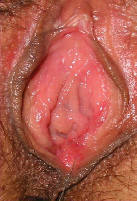
Figure 7.1 Vulvar papillomatosis widely involving the vestibule, a common nonpathological condition. Its bilateral and symmetrical array helps differentiate this normal vulvar feature from vestibular condyloma acuminate.

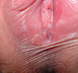
The mucosa of the labia minora and vestibule is generally pink and smooth; however, localized or widespread micropapillary or villiform patterns may sometimes be observed (Figure 7.1).
These occasionally coalesce to form tissue that can be misinterpreted as condyloma due to the human papillomavirus (HPV) virus. These papillae can be distinguished from HPV-induced lesions on the basis of their regular shape and distribution, uniform color, soft consistency, and lack of tendency to fuse. They are projections of connective tissue covered by a normal epithelium. In contrast, condylomas are pointed, soft, pink, or white finger-elongated excrescences (Figure 7.2). Prominently vascularised, they have fingerlike projections on the surface; their appearance differs according to the site affected. On hair-bearing skin, they are flesh-colored and somewhat camouflaged; on hairless skin, they tend to be soft, papular,and strikingly white; on mucous membranes they are often fleshy, vascular, and filiform. While these vulvoscopic findings of the morphology of condylomata are quite specific, a vulvar biopsy can confirm the diagnosis and rule out VIN. In addition, polymerase chain reaction (PCR) can be performed on the biopsy to determine the strain(s) of HPV present.
Vulvoscopic Finding of Vulvodynia
The International Society for the Study of Vulvovaginal Disease (ISSVD) defines vulvodynia as vulvar discomfort occurring in the absence of relevant visible findings [4]. Allodynia and hyperalgesia are usually present, and are evidenced by discomfort from spontaneous and/or provoked pain. Dyspareunia is not always present, but most patients do have discomfort during sexual intercourse. Patients frequently complain of pain involving the introitus (vestibulodynia) or pain radiating to the labia majora, groin, and perineum (generalized vulvodynia). Most of the time, the only finding during the vulvoscopic examination of a women with vulvodynia is erythema of the vestibule, which is frequently most apparent at the ostia of the major and minor vestibular glands and Skene’s glands. Therefore, the vulvoscopist’s main goal in a woman with suspected vulvodynia is to rule out specific diseases that can cause vulvar pain (Figure 7.3).
Figure 7.3 Vestibulodynia. The vulvoscopy shows no abnormality other than some degree of vestibular erythema.
Vulvoscopic Finding of Ulcerative Conditions
Related posts:


Stay updated, free articles. Join our Telegram channel

Full access? Get Clinical Tree


