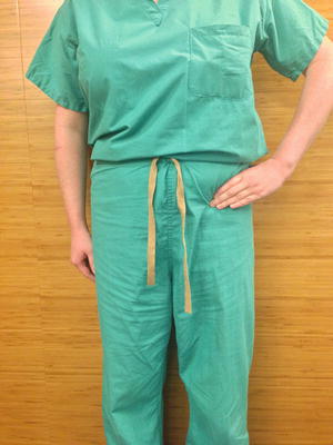
Fig. 17.1.
Intra-articular hip pathology is excluded by painless hip rotation and a negative Stinchfield test.
Lumbar Spine Disease
Spine diseases, such as thoracolumbar discogenic pain at multiple levels or L2–L4 nerve root impingement, can all cause groin and thigh pain, with or without associated back pain [4]. The mechanism is sometimes complex, as the pain can be radicular pain from nerve root compression or pain from nerve endings on the herniated disc itself. The existence of sensory nerve endings in the annulus fibrosus of the human lumbar intervertebral disc has been described and well documented [5]. Patients sometimes find it hard to understand how their groin pain is originating from their back when they do not present with back pain. Physical exam of the hip is usually normal. Limited spine flexion, extension, and/or lateral bending are sometimes seen. Many of these patients have very weak core musculature. The femoral nerve traction test is done with the patient in a prone position with the knee flexed to 90° and the hip fully extended; pain in the anterior thigh suggests a L2–L4 nerve root impingement. One of the most common findings, however, appears to be tight hamstring musculature. Tight hamstrings lead to hip flexion contractures, a subtle crouched gait, and compensatory pressure on the spine. Physical therapy focusing on core strengthening, hamstring stretching, and lumbar stabilization is the first line of treatment. MRI of the spine is sometimes needed, followed by selective spinal diagnostic and therapeutic injections.
Osteitis Pubis
Osteitis pubis is a noninfectious inflammatory process of the pubic symphysis commonly seen in runners and in athletes involved in cutting sports such as soccer and hockey. Previous trauma, overuse, and vaginal delivery are all risk factors. Patients often present with groin pain that is activity related. On physical exam they usually have normal hip range of motion, nontender abductor muscles laterally, and focal tenderness to palpation over the pubic symphysis. Weak core musculature is often noted; pain with resisted hip adduction or passive hip abduction may also be found. These latter physical exam findings are often confirmed by tendinosis of the rectus abdominis and adductor longus insertions on an MRI. Plain radiographs of the pelvis are usually helpful as well, as they typically show widening of the symphysis with blurring of the cortical margins and sometimes cysts. Physical therapy is the first line of treatment followed by steroid injections. Surgical bony resection with preservation of the rectus and pubic ligaments is rarely done [6].
Pubic Ramus Fractures
Fractures of the superior and/or inferior pubic rami are commonly seen in elderly patients who have sustained a low-energy fall. These patients present with acute groin pain that was not present prior to trauma. They usually have painless hip rotation but focal tenderness over the bony pelvis lateral to the pubic symphysis. It is common for these patients to report having had recent hip radiographs that were normal. Unfortunately, the pelvis has rarely been evaluated. Hip radiographs often do not show rami fractures; it is imperative to obtain an AP of the pelvis when evaluating groin pain. Treatment of these fractures is most commonly nonoperative with unrestricted weight bearing.
Iliopsoas Pathology
The two most common iliopsoas pathologies seen by orthopedic surgeons are snapping (coxa saltans interna) and tendinitis. Internal snapping of the iliopsoas tendon is actually an extra-articular process. Patients usually present with an audible snap and anterior groin pain. As the hip is extended, the iliopsoas tendon travels from lateral to medial catches at the iliopectineal eminence or on the femoral head. On occasion, the snapping can be palpated directly in the groin. Dynamic ultrasound may be useful in the diagnosis. MRI is sometimes indicated, as it can show resultant hip labral tea [7]. Iliopsoas tendinitis is a relatively rare entity in patients with native hips and seen most commonly in patients involved in activities that require repetitive hip flexion (rowing, uphill running, and ballet). Patients generally present with anterior hip pain that radiates to the knee and sometimes with knee pain alone. The most common physical exam findings are painless hip rotation, pain with resisted hip flexion, and pain with passive hip extension. Initial treatment of both coxa saltans interna and iliopsoas tendinitis is always stretching (best done in a luge position) and, when necessary, steroid injections [8]. Open and arthroscopic releases of the iliopsoas tendon have been reported, but complications such as symptomatic intra-abdominal fluid extravasation [9] and anterior hip instability [10] have been reported. Exclusion of the other potentially life-threatening pathologies with which abdominal surgeons are very familiar, such as an iliopsoas abscess, is clearly important.
Intra-articular Causes of Groin Pain
Anterior groin pain that radiates deep into the hip and sometimes radiates into the groin should always raise the suspicion of an intra-articular process. In the now classic C sign (Fig. 17.2) suggestive of intra-articular hip pathology, patients will place their hand over the affected hip with the index finger in the groin and the thumb placed proximal to the greater trochanter in the shape of the letter C [11, 12]. Patients will also commonly have a positive Stinchfield test, described earlier in this chapter. Lastly, an active straight leg raise with the supine patient actively raising the heel of the leg by flexing the hip about 30° is also suggestive of intra-articular pathology: during this test, hip flexors produce joint reactive forces up to two times the patient’s body weight across the hip joint itself.


Fig. 17.2.
The classic C sign suggestive of intra-articular hip pathology.
Arthritis and Avascular Necrosis
More than 21 % of the US population aged 18 or older have arthritis or other rheumatic conditions, and that percentage increases as people age. The number of people in the USA who have arthritis is projected to increase to 67 million, or 25 % of the adult population, by the year 2030. Osteoarthritis is the most common form and the hip is commonly affected. Patients present with pain in the groin, diminished hip motion, difficulty putting on their shoes or socks, and inability to ambulate extensively. Physical exam reveals a positive Stinchfield test and C sign. Nonoperative treatment consists of intra-articular steroid or hyaluronate injections [13]. Surgical intervention is a total hip arthroplasty. Avascular necrosis, commonly seen in the femoral head, is also a common reason for groin pain, especially in patients with risk factors such as steroid use, alcohol abuse, coagulopathies, sickle cell disease, Gaucher’s disease, and decompression sickness. When radiographs are normal and suspicion is high, patients should undergo an MRI of the hip. Depending on the stage of avascular necrosis, treatment includes protected weight bearing, bisphosphonate treatment, electrical stimulation, electromagnetic fields, core decompression, bone grafting, autologous mesenchymal cells, osteotomies, and arthroplasty procedures [14].
Hip Synovitis and Septic Arthritis
Transient synovitis of the hip is a short-lived acute inflammatory process usually seen in boys aged 2–10 following an upper respiratory tract infection. Generally a diagnosis of exclusion, it must be differentiated from a septic hip, which is also commonly seen in this patient population. Patients present with groin pain and sometimes difficulty putting weight on the limb. In addition to the aforementioned tests for intra-articular pathology, these patients will have pain with log rolling of the hip while in extension. Kocher et al. have provided useful ways of differentiating between these two entities [15]. Patients with transient synovitis require close observation, while those with septic arthritis most commonly require arthroscopic or open irrigation and debridement of the hip joint.
Femoroacetabular Impingement
Femoroacetabular impingement (FAI) occurs when anatomic variations in hip anatomy lead to impingement between the acetabulum and the femoral head–neck junction. FAI is believed by many to be a common pathway to hip arthritis, especially in younger patients. Impingement is generally classified into Cam impingement and Pincer impingement. CAM impingement is due to prominence of the anterosuperior head–neck junction or diminished head–neck offset. Pincer impingement is secondary to acetabular overcoverage of the femoral head for a variety of reasons such as coxa profunda or acetabular retroversion. FAI may lead to chronic groin pain, especially in younger adults who often go on to have symptomatic arthritis. FAI is also a probable predisposing factor to labral tears, most of which, even in the face of trauma, would probably rarely occur otherwise. Patients with FAI may have some of the classic signs of intra-articular hip pathology and also report pain with tests such as the anterior impingement test or FADIR (flexion adduction internal rotation). Sophisticated radiographs and MR arthrography are some of the methods of choice in further studying these conditions [16]. Diagnostic hip injections, when positive, may lead to a variety of arthroscopic and/or open joint preserving procedures [17].
Stay updated, free articles. Join our Telegram channel

Full access? Get Clinical Tree


