Fig. 41.1.
Osteitis pubalgia on MRI.
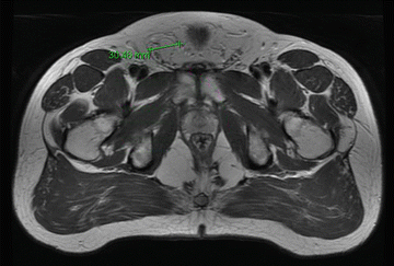
Fig. 41.2.
Right inguinal hernia on MRI.
Diagnosis
While the reducible right inguinal hernia in this patient was obvious, the patient’s history and physical examination gave high suspicion for the clinical diagnosis of osteitis pubis as well. The symptoms of osteitis pubis can be presented as any complaint in the groin or lower abdomen [1]. Pain generally is localized over the symphysis and may radiate to the groin, scrotum, perineum, medial thigh, hip, or abdomen [2].
Physical findings for osteitis pubis can vary. It is important always to consider any sport involved, as well as the chronicity of the conditions [3]. Gradual onset of pelvic pain, with pubic symphysis tenderness, is one of the features of osteitis pubis [4]. One great modality for the diagnostic investigation in detecting the osteitis pubalgia is MRI. The differential diagnoses of chronic groin pain that can present similarly to osteitis pubis include musculotendinous strain, osteomyelitis, inguinal hernia, referred low back pain, intra-articular hip disease and genitourinary disease, and adductor tendinopathy. Osteomyelitis of the pubic symphysis is one of the diseases most commonly confused with osteitis pubis [5], so clinical evaluation requires a careful synthesis of history taking, physical examination, and appropriately directed investigation.
Discussion
Osteitis pubis, a rare condition, is characterized by pelvic pain localized over the symphysis pubis, in the lower abdominal muscles or in the perineum. The pain may radiate to the adductor region of the thigh, and patients may describe painful adductor muscle spasms. Aggravating factors are walking and standing from a seated position.
Osteitis pubis, also known as pubalgia or sometimes mislabeled as just athletic pubalgia, is one of the most chronic and debilitating syndromes affecting athletes [6]. It is described as the pubic bone stress injury occurring usually as a result of chronic overload or impaction trauma [7].
Osteitis pubis is also considered as noninfectious, inflammatory damage to the pubic symphysis and its supporting structures [8] anatomically. The pubic symphysis is mainly composed of fibrocartilage and is a nonsynovial, nonvascular joint. The pubic symphysis is reliant on four ligaments to maintain its supportive integrity. Most of the strength and support arise from the superior and inferior ligaments, whereas the anterior and posterior ligaments are of less supportive importance. The pelvic floor musculature, composed of the levator ani and coccygeus, inserts posteriorly at the pubic symphysis. The pectineus, rectus abdominis, and oblique externus muscles, as well as the inguinal ligament, insert near the superior portion of the pubic symphysis. The pubic rami give rise to several muscle origins: adductor magnus, adductor longus, adductor brevis, and gracilis. These muscles make up the adductors of the hip [1].
The prevalence of osteitis pubis among the general population of athletes ranges from 0.5 to 6.2 % [8, 9]. Although many different sports may be associated with osteitis pubis, sports with a higher risk include soccer, football, ice hockey, and rugby [9].
The etiology of osteitis pubis is unknown; repetitive trauma alone or in conjunction with opposing shearing forces across the pubic symphysis is likely the main contributing factor in many athletes [10, 11]. This may be due to different types of movements, including rapid acceleration-deceleration, kicking, and changes in direction.
Management
Nonoperativemanagement of osteitis pubis is similar to that of other causes of chronic groin pain and consists mainly of rest, ice or heat, and nonsteroidal anti-inflammatory drugs (NSAIDs) or other oral medication if patient can not take an anti-inflammatory medication. If these initial modalities are not helpful after a defined trial period (3 weeks for example), then the next intervention to consider would be glucocorticoid injections directly into the pubic symphysis or oral glucocorticoids [12, 13]. Additional nonoperative interventions can then also include physiotherapy focusing on core stability, muscle balance, and rotational hip range of movement; and activity modification. The most important lesson here is that if a patient has osteitis pubalgia and an inguinal hernia, just repairing the hernia alone will not alleviate the pubalgia pain. We therefore recommend initiating treatment for pubalgia first and delaying a hernia repair until the pubalgia pain is better controlled and the patient understands the difference between the hernia pain and the pubalgia pains.
Related posts:
 Introduction to Primary and Secondary Groin Pain: What Is Inguinodynia?
Prevention of Pain: Optimizing the Laparoscopic TEP and TAPP Techniques
Prophylactic Neurectomy Versus Pragmatic Neurectomy
Patient with Referred Hip Pain
Introduction to Primary and Secondary Groin Pain: What Is Inguinodynia?
Prevention of Pain: Optimizing the Laparoscopic TEP and TAPP Techniques
Prophylactic Neurectomy Versus Pragmatic Neurectomy
Patient with Referred Hip Pain
 Chronic Pelvic Pain in Women
 Groin Pain Etiology: Hip-Referred Groin Pain
Stay updated, free articles. Join our Telegram channel

Full access? Get Clinical Tree


