Extension
20°
Flexion
>120°
Adduction
20°
Abduction
40°
Internal rotation hip in extension
30°
Internal rotation hip in flexion at 90°
20°
External rotation hip in extension
50°
External rotation hip in flexion at 90°
30°
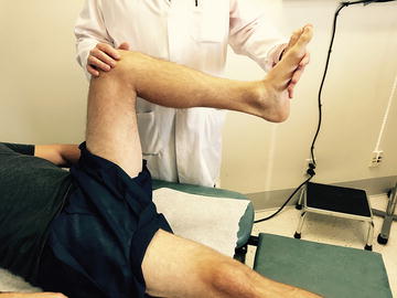
Fig. 8.1.
The physical examination maneuver to determine range of motion about the hip. The extent of flexion of the hip is assessed in neutral rotation.
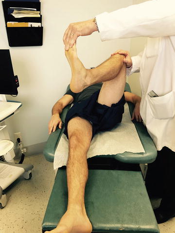
Fig. 8.2.
The extent of external rotation of the hip is assessed with the hip in 90 degrees of flexion.
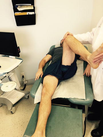
Fig. 8.3.
The extent of internal rotation of the hip is assessed with the hip in 90 degrees of flexion.
When thinking about groin pain referred from the hip, it is helpful to think of the differential as problems related to either the soft tissue or skeleton. Further separating these into architectural versus physiological causes helps to clarify thinking. Although considerable overlap occurs with many processes, it remains a useful analytical framework. For the sake of brevity, trauma will be excluded from the discussion; however, suspicion of fracture following even minor trauma should remain high. Any fracture of or about the hip or pelvis should be treated with protected weight bearing and immediate referral to an orthopedic surgeon.
Groin Pain from the Bone
Architectural Problem
Osteoarthritis
Presentation
Osteoarthritis (OA) is extremely common in an aging patient population. It is estimated to affect 60 million Americans by the year 2020 [5]. In any patient over 50 years, it should be high on one’s differential as the cause of groin pain. Most patients with OA present with a slowly progressive pain in the groin, hip, or thigh, typically worse in the morning and at night. Patients describe an aching pain that improves with light activity and is worse with strenuous activity. Patients commonly report difficulties with initial motion after prolonged periods of rest, with improvement after a few steps. More advanced hip OA eventually results in stiffness and difficulty with activities of daily living.
Physical Exam
The classic finding of crepitus with range of motion is rare. Patients may present with a very stiff joint, often with back pain that is more severe than the hip pain. Alternatively, some patients present with severe pain on weight bearing, with an almost normal range of motion. An important and early clinical sign of hip OA is decrease in internal rotation. Passive external rotation during flexion of the hip is known as Drehmann’s sign and is indicative of this loss of internal rotation. Eliciting a positive Stinchfield test , which results in pain at the hip with resisted straight-leg raise, is sensative however has low specificity (Fig. 8.4).
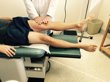
Fig. 8.4.
The Stinchfield test. The patient performs a forced straight-leg raise against downward resistance at the thigh placed by the examiner. Pain in the groin with this maneuver is considered positive.
Diagnostic Exams
The most useful radiological study is a standing low anteroposterior (AP) pelvis (including both hips) with the patient bearing weight equally on both sides (Fig. 8.5). A supine lateral radiograph of the affected side (“frog leg lateral”) will complete the examination. These two simple views will help to elucidate more than 90 % of hip-referred groin pain originating from the bone. OA will have an obvious appearance on plain x-rays, and no further imaging is needed to arrive at this diagnosis (Fig. 8.6).
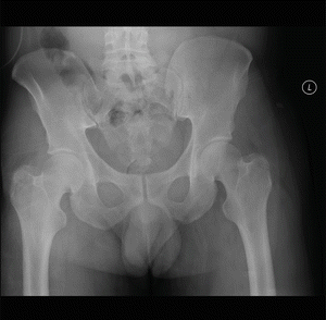
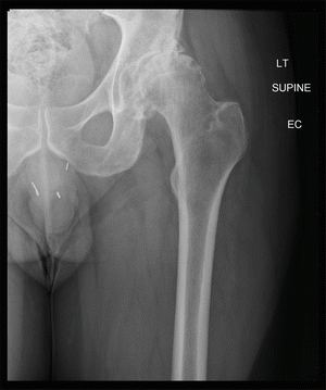
Fig. 8.5.
Radiograph of a normal pelvis.
Fig. 8.6.
Radiograph of the left hip showing changes typical of advanced osteoarthritis. Note joint space narrowing, subchondral sclerosis, osteophyte and cyst formation in the femoral head and acetabulum.
Differential
Patients, especially young ones, with no obvious OA but bony abnormalities should receive a consultation with an orthopedic surgeon. Some of these pathologies can be treated early (e.g., impingement or hip dysplasia), leading to decreased rates of degeneration about the hip.
Appropriate Treatment/Referral
Osteoarthritis is very common, and its first line of treatment is simple: nonsteroidal anti-inflammatory drugs (NSAIDs) , stretching, physical therapy (including pool therapy ), and weight loss. Once these treatments have been exhausted, the patient should probably be referred to a specialist. Steroid injection under ultrasound or other imaging modalities should be considered if the pain is very acute. Hip arthroplasty is an effective and reproducible procedure but should be carefully weighed against the risks of the procedure. Finnish Registry data suggest a 15-year revision-free survival rate of 71–86 % for total hip arthroplasty [6]. Not unlike other complex procedures, outcomes are better when performed by high-volume surgeons in high-volume centers.
Femoroacetabular Impingement
Presentation
Femoroacetabular impingement (FAI) is a developmental abnormality of either the femoral head-neck junction and/or the acetabulum, either of which leads to abnormal hip function. These patients are generally young and/or active. They fall into two broad categories: cam-type impingement (loss of femoral head-neck offset) and pincer-type impingement (acetabular over-coverage). These biomechanical abnormalities lead to tears of the acetabular labrum (discussed in the next section) and delamination of the cartilage. This is theorized to be the precursor of the so-called idiopathic OA; however, it is not yet clear if surgical intervention has any influence on development of OA later in life [7].
Physical Exam
Groin pain with anterior impingement is exacerbated with high flexion, adduction, and internal rotation at the hip (Fig. 8.7). Alternatively, posterior impingement is made worse with extension and external rotation [1]. Either of these may be combined with or exclusively present with labral-type symptoms, often with a popping and catching sensation with motion, which causes pain.
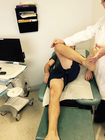
Fig. 8.7.
The impingement maneuver consisting of flexion, adduction, and internal rotation. Pain or a pinching sensation in the groin during this maneuver is considered to be indicative of impingement.
Diagnostic Exams
Low AP weight-bearing pelvis (including both hips) with a supine lateral radiograph of the affected side (“frog leg lateral”) is recommended. Radiographic measurements are taken to assess for these abnormalities, as they are often subtle (Fig. 8.8).
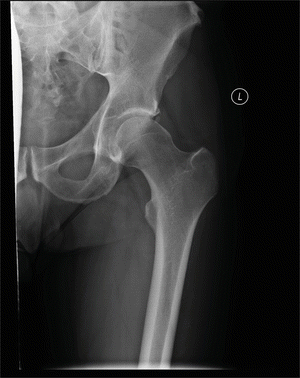
Fig. 8.8.
Anteroposterior (AP) radiograph of the left hip showing the typical cam deformity of the proximal femur with os acetabulum (an accessory bone unrelated to the pathology).
Differential
Cam-type impingement is classically described among young athletic males. Given this population, it is important to rule out muscular strain or even sports hernia . Femoral hernia should be considered among women, even if pincer-type impingement is noted. The strict definition of cam versus pincer type impringement is somewhat of an oversimplification, however, with as much as 80 % of cases being considered a combined mechanism [9].
Appropriate Treatment/Referral
Both open and laparoscopic treatments have been effective in the control of symptoms. If FAI is suspected on clinical or radiographic examination, referral to an orthopedic surgeon who has experience with FAI is important, as early intervention may delay or prevent progression of the degeneration of the joint.
Labral Tear
Presentation
The acetabular labrum has been shown to have a role in maintaining appropriate synovial fluid pressure for adequate lubrication of the hip joint [3]. The best analogy is a rubber gasket in a hydraulic joint. As such, the labrum has received new attention regarding its potential role in preserving the hip cartilage. Patients with tears of the labrum often present with deep-seated hip or groin pain or report a popping or clicking sensation with motion.
Physical Exam
Painful range of motion is present, most pronounced with flexion or extension of the hip in abduction, combined with a rotational movement. Rolling the hip through this range of motion often produces pain and a popping sensation for a patient with labral pathology.
Diagnostic Exams
X-rays may occasionally show a small calcification at the acetabular rim, indicating a calcified labrum from recurrent trauma and degeneration. However, most labral pathologies are not diagnosed with plain radiographs. Magnetic resonance (MR) arthrogram is the imaging study of choice for diagnosis and when a hip joint preservation procedure is a consideration (Fig. 8.9).
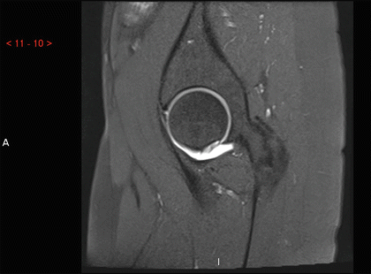
Fig. 8.9.
T2-weighted MR arthrogram showing contrast tracking between the acetabular rim and the labrum indicative of a labral tear.
Differential
Up to 90 % of patients with labral pathology have some degree of FAI according to some authors [7]. As such, an evaluation should include radiographic measurements, including the alpha angle and center edge angle to evaluate for this entity.
Appropriate Treatment/Referral
Referral to an orthopedic surgeon with experience treating FAI is indicated due to the common coexistence of these pathologies. Although not an emergency, early intervention may decrease long-term degeneration of the joint.
Hip Dysplasia
Presentation
Whereas symptoms of acetabular impingement occur due to actual or effective over-coverage of the femoral head, hip dysplasia represents the other side of this spectrum: under-coverage of the femoral head leads to increased stresses on the chondral surfaces. Congenital hip dysplasia in its extreme form will lead to dislocation of the hip among infants; however, the disease process lies on a spectrum, and many patients with dysplastic hips may be asymptomatic for many years prior to diagnosis. Additional conditions such as spondyloepiphyseal dysplasia and achondroplasia commonly lead to malformations of the hip that lead to increased rates of osteoarthritis. As such, any patient affected with dwarfism should be evaluated for orthopedic causes with any presentation of groin pain.
Physical Exam
Painful range of motion, positive Stinchfield test (see the section on Osteoarthritis above), and classic signs of early osteoarthritis are present among these patients (see Fig. 8.4).
Diagnostic Exams
Plain x-rays are often adequate to diagnose both the architectural problems and any early degenerative process occurring of the joints. However, patients without radiographically obvious degenerative changes but clinically evident hip dysplasia should continue to receive further orthopedic evaluation.
Appropriate Treatment/Referral
As hip dysplasia is a congenital problem, patients with this malformation often present at relatively young ages with the beginning of degenerative changes. If caught early enough, operative options exist to increase the coverage of the femoral head with periacetabular osteotomies that may lead to decreased rate of degeneration of the hip. Once a patient has progressed to advanced degeneration of the joint, the only effective option is joint arthroplasty, which, although successful in pain relief and restoration of more normal biomechanics, has a limited life span. Revision surgery has much less reliably positive results and a higher complication rate. Referral to an orthopedic surgeon with experience with these procedures is important to delay progression as long as possible.
Occult Fracture
Presentation
Although it is uncommon for a patient to initially present to the doctor’s office with a hip fracture, it is possible that a patient may have had a prior workup that was falsely negative and is now presenting with groin pain that is in fact due to a missed hip fracture. This scenario may be seen among patients who suffered a trauma or fall with no clear x-ray evidence of a fracture. Although most emergency departments or urgent care centers will adequately work up a nondisplaced fracture seen on x-ray with computed tomography (CT) scan or MRI, an occult hip fracture is an important diagnosis to consider among patients who have a history of trauma or fall and pain, but with no obvious x-ray evidence of fracture. Nondisplaced fractures of the femoral neck, pubic rami, and sacrum are common following falls in elderly patients.
Physical Exam
Patients present with groin and hip pain, typically exacerbated with any movement about the hip. Among patients with pubic ramus fractures, palpation of the pubic symphysis is often particularly painful. It is also important to palpate the sacrum, as tenderness to palpation may represent a fracture of the sacral ala.
Diagnostic Exams
Among those with nondiagnostic x-rays, CT scan will help demonstrate nondisplaced and minimally displaced fractures about the hip. However, MRI is preferred, specifically for femoral neck fractures, as it is 100 % sensitive in the detection of radiographically occult femoral neck fractures [8]. A black line within the bone on T1-weighted images indicates a nondisplaced fracture.
Appropriate Treatment/Referral
Nondisplaced pubic ramus and sacral ala fractures may be treated with simple pain control and radiographic follow-up to ensure that no unrecognized instability is present. If x-ray examination remains stable following mobilization, the patient does not require protected weight bearing; however, a walking aid should be recommended to ensure stability. On the other hand, nondisplaced femoral neck and intertrochanteric fractures require strict non-weight bearing and immediate referral to an orthopedic surgeon, as displacement may lead to a more difficult surgical treatment or displacement of the fragment.
Physiological
Septic Hip
Presentation
A septic joint typically presents with acute onset hip and/or groin pain that is exacerbated by movement. Patients may or may not demonstrate erythema and swelling, due to the extent of the soft tissue surrounding the hip. A history of recent sexual contacts should be obtained among those who are sexually active, as gonococcal infections are known to present with monoarticular septic joints. Consideration of this diagnosis should also be considered among immunocompromised patients.
Physical Exam
Patients report a painful joint, with dramatic increase in pain with any motion. It is this sign of irritable range of motion, with even small movements, that is the most reliable of the clinical signs. Fevers, chills, and leukocytosis may be seen; however, their absence does not exclude the diagnosis of a septic hip.
Diagnostic Exams
X-rays most often do not show any abnormalities, with the exception of long-standing cases of osteomyelitis in which a sequestrum and involucrum have had time to evolve. MRI can be useful if considering osteomyelitis. However, in the case of a simple septic joint, MRI will provide no more information other than the presence and size of the effusion. Aspiration and targeted surgical and antibiotic treatment are used as the standard of treatment. Synovial fluid is routinely sent for crystal examination, cell counts, and cultures. Cell counts above 50,000 white blood cells (WBC) and with greater than 75 % polymononuclear (PMN) cells are considered indicative of infection and are typically taken to the operating room for joint irrigation and debridement [9]. It is important to note that immunosuppressed patients may have an infected joint space in the presence of lower cell counts. Culture examination, although a critical part of the examination, does not trump the need for operative decompression and irrigation of the septic hip.
With any prosthetic joint in which infection is a concern, erythrocyte sedimentation rate (ESR) and C-reactive protein (CRP) need to be checked in addition to a complete blood count (CBC). ESR above 30 mm/h and of CRP greater than 1 mg/dL should raise suspicion for prosthetic joint infection. As in native hips, aspiration of the joint with cell counts is the gold standard for diagnosis, however cell counts as low as 1760 cells/μL are suggestive of a periprosthetic infection [2].
Differential
It is important to consider crystalline arthropathy in the differential diagnosis of a septic joint, as its clinical presentation is nearly indistinguishable from that of a septic joint. Additionally, even if crystals are seen on joint aspiration, in the scenario of high number of WBCs and a high percentage of PMNs, the physician should consider the possibility of a superimposed infectious process. Cultures should be followed for a minimum of 3 days to rule out this possibility. However, if the suspicion is high for an infection, one should expeditiously proceed with operative treatment, i.e., decompression, irrigation, and debridement of the infected joint.
Related posts:
 Introduction to Primary and Secondary Groin Pain: What Is Inguinodynia?
Prevention of Pain: Optimizing the Laparoscopic TEP and TAPP Techniques
Prophylactic Neurectomy Versus Pragmatic Neurectomy
Patient with Referred Hip Pain
Introduction to Primary and Secondary Groin Pain: What Is Inguinodynia?
Prevention of Pain: Optimizing the Laparoscopic TEP and TAPP Techniques
Prophylactic Neurectomy Versus Pragmatic Neurectomy
Patient with Referred Hip Pain
 Chronic Pelvic Pain in Women
 Radiologic Evaluation for Postoperative Groin Pain
Stay updated, free articles. Join our Telegram channel

Full access? Get Clinical Tree


