Fig. 19.1.
Axial MR T1-weighted, T2-weighted, and fat-saturated T2-weighted images of bilateral flat mesh repairs. The normal mesh on the left (white arrow) appears smooth and linear, particularly on T1. Dark “blooming” artifacts (curved white arrow) are commonly encountered in the postoperative groin and are due to the presence of metal tacks or staples. In comparison, the mesh on the right is distorted and difficult to visualize (black arrow) due to mesh infection. There is a large fluid collection (curved black arrows) just posterior to mesh material, best visualized on the fluid-sensitive T2 sequences.

Fig. 19.2.
Axial MR T1-weighted, T2-weighted, and fat-saturated T2-weighted images of bilateral flat mesh repairs. Both mesh are intact; however, large superficial fluid collections are seen bilaterally (black arrows). While simple seroma would appear bright on T2 sequences only, this fluid is also intermediately bright on T1 sequences, suggesting the presence of significant blood products. Both groins were percutaneously drained, demonstrating hematoma.
Mesh Complications
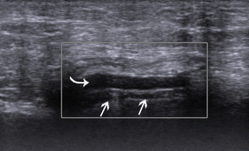
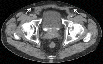
Mesh complications often present with subtle imaging findings, and may require knowledge of the operative technique utilized to diagnose definitively. US does not reliably identify the mesh, especially if it is folded, balled up, or otherwise complicated (Fig. 19.3). As such, US is not recommended as a first-line imaging modality to evaluate the postoperative groin after mesh implantation when the integrity of the mesh itself is in question. Due to the combination of low material density and minimal profile, normal mesh material is often indistinguishable from surrounding tissue on CT [2], requiring the radiologist to discern a postoperative state from the presence and location of the patient’s surgical scars (Fig. 19.4). Even in states of chronic inflammation (e.g., mesh reaction), it may be impossible to specifically identify pathology on the basis of CT alone. On MR, flat mesh materials appear as dark linear bands on T1 sequences, slightly thicker than normal fascial planes, but may be more difficult to identify among their surrounding tissues on fluid-sensitive sequences (Fig. 19.5).


Fig. 19.3.
The flat mesh (white arrows) shown on this Doppler US is hardly conspicuous, and would be even less so if not for the small fluid collection (curved white arrow) overlying it.

Fig. 19.4.
The bilateral flat mesh (white arrows) seen in this axial CT of the pelvis look similar to scar tissue, making it difficult to differentiate subtle mesh abnormalities.

Fig. 19.5.
Coronal MR T1-weighted, axial T1- weighted, and axial fat-saturated T2-weighted images of flat mesh material (black arrows) closing an indirect defect of the right inguinal canal (white arrow). Flat mesh materials typically appear as a thick hypointense line. While there is some undulation of this mesh, there is no inappropriate folding and no significant mass effect to suggest meshoma or mesh reaction.
Normal mesh should appear smooth, and wrinkling may represent migration with subsequent focal recurrence or inflammatory response. While mesh may be fixed in position with sutures, staples, or tacks, shifting of mesh material may occur. If slippage does occur, even partial mesh migration may result in gross recurrence of bowel protrusion, or may present as subtle herniation of peritoneal or preperitoneal fat. Interposition of even small amounts of fat between mesh material and the abdominal wall may be a cause of significant pain. Dynamic MR sequences are particularly capable of identifying such pathology, which may be missed by CT.
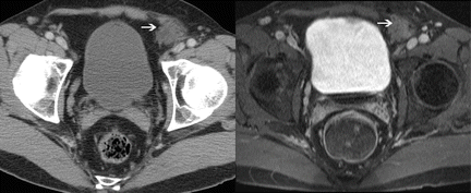
Pain associated with more complex mesh materials such as plug or sandwich designs are uniquely challenging to diagnose, as many radiologists are not aware of the existence of such materials and may not be able to recognize normal postoperative appearance without access to detailed operative notes or direct communication by the referring physician. Volume-occupying materials utilized in repair of large, patulous defects often incorporate significant biological material into their interstices, appearing on imaging as large pseudomasses and resulting in misdiagnosis (Fig. 19.6). As an example, our own personal case series includes a post-herniorrhaphy patient with significant pain who underwent percutaneous biopsy of “bilateral inguinal masses” that were in fact mesh PerfixTM plugs that were impressing upon the contents of the inguinal canal, resulting in testicular venous congestion and chronic pain. Most plugs are found within the inguinal canal with a cone appearance, and conversion to a different shape (especially a narrow needle-like one) may be indicative of abnormality, suggesting migration as an etiology of groin pain. When properly positioned, sandwich mesh, such as the ProleneTM Hernia System , should be seen on MR as a T1 dark band superficial to the internal inguinal ring or direct fascial defect, with a second layer positioned preperitoneally: the presence of fat between the layers represents normal appearance.


Fig. 19.6.


Contrast-enhanced axial CT and corresponding contrast enhanced T1-weighted axial MR images of mesh plug within the left direct space. Both modalities reveal a large “mass” (white arrows) with similar density/intensity as soft tissue. This appearance is due to incorporation of soft tissue within the mesh plug’s interstices. Though there is mild irregularity within the plug, the lack of contrast enhancement and normal appearance of the surrounding fat confirms lack of mesh pathology.
Stay updated, free articles. Join our Telegram channel

Full access? Get Clinical Tree


