The Nervous System


The Nervous System


INTRODUCTION

The central nervous system (CNS) alterations that are common in surgical critical illness are listed in Table 9.1. The most common one is traumatic brain injury (TBI). A description of basic brain anatomy and cerebral oxygenation physiology serves as a practical underpinning to the understanding and management of several of these alterations.


Anatomy

Knowledge of the anatomy of the cerebral cortex, with sensory and motor loci, is important for assessing focal sensory and motor deficits. However, many etiologies of depressed mental status in surgical critical illness (e.g., diffuse axonal injury, brain hypoperfusion, narcosis) produce global alterations in cortical function that may or may not be associated with localized findings. Important for assessing altered mental status is an understanding of the anatomy of the reticular activating system (RAS), a diffuse group of neurons that extend along the central brainstem from the medulla to the thalamus (Fig. 9.1). RAS is stimulated by every major somatic and special sensory pathway and serves to activate the cerebral cortex. Since the RAS arises in the brainstem, knowledge of the anatomy of the brainstem is useful for evaluating the status of the RAS.

The brain is enclosed in a hard, rough chamber, which, while being protective, can be responsible for direct or “counter coup” injury (Fig. 9.2). The medial temporal lobe is positioned close to the midbrain, the path of the third cranial nerve and cerebral blood vessels (Fig. 9.3). Particularly, medial displacement of the temporal lobe is likely to interrupt first the function of the third cranial nerve, then the midbrain, and, subsequently, larger areas of the brainstem (Fig. 9.4).

The brainstem is divided into midbrain, pons, and medulla (Figs. 9.5 and 9.6). Brainstem nerves that are valuable to evaluate include the following: (1) the third for innervation of the medial rectus and parasympathetic innervation to the pupil; (2) the fifth for sensation to the cornea; (3) the seventh for motor to the eyelids; (4) the sixth for innervation of the lateral rectus; (5) the eighth for innervation of the vestibular apparatus; (6) the medial longitudinal fasciculus connecting the eighth to sixth and third; (7) the course of the sympathetic nervous system through the entire brainstem.

Consciousness requires cortical function and is not lost unless cortical function is diffusely diminished. Diffusely diminished cortical function may result either from direct, global disruption of cortical function or from disruption of the RAS in the brainstem. Since the cortex is more sensitive to metabolic disturbances than the brainstem (i.e., hypoglycemia, hypoxia), “cortical coma” is more likely to be “metabolic,” whereas “brainstem coma” most often is secondary to pressure on or structural damage to the brainstem (1).


Physiology of Cerebral Blood Flow, Oxygen Metabolism, and Intracranial Pressure

The brain requires a continuous supply of oxygen and glucose to support the aerobic glycolysis necessary to maintain the integrity of brain neurons. The brain is proportionally more sensitive to decreased delivery of oxygen than decreased delivery of glucose. The cerebral metabolic rate of oxygen (CMRO2) is a useful measure of brain metabolism and is calculated as follows:


CMRO2 = CBF × C(a-v)O2

Where, CBF = cerebral blood flow; C = oxygen content; a, v = arterial, venous, respectively.

In adults who are awake, CBF approximates 50 ml/100 g tissue at PaCO2 of 40 mm Hg. CMRO2 is about 3.2 ml/100 g under these conditions. CMRO2 may increase with activities such as seizures and decrease with drug-induced coma. Usually, CBF adjusts to meet alterations in
CMRO2. However, severe brain injury may lead to disruption of the autoregulation of CBF such that too much or too little CBF may be supplied for the metabolic demands of the brain.


Table 9.1 Central Nervous System Alterations in Surgical Critical Illness


  1. Head trauma


    1. Pathophysiology of brain injury


    2. Examination before and after the CT scan


    3. Management of intracranial and cerebral perfusion pressure


    4. Adjuncts for management


    5. Associated organ malfunction


  2. Blunt cerebrovascular injury


  3. Manifestations of spinal cord injury


  4. Neuromuscular disorder of surgical critical illness


  5. Management priorities for patients with traumatic brain injury


Figure 9.1 A schematic representation of the reticular activating system arising from the brainstem and projecting impulses cephalad throughout the cortex. Source: ACS. ATLS Student Manual. Chicago: ACS, 1997.

CBF is primarily determined by cerebral perfusion pressure (CPP), which is the difference between mean arterial pressure (MAP) and intracranial pressure (ICP), as well as the arteriolar vascular resistance in the brain tissue. Therefore, CBF will increase if CPP increases and/or arteriolar resistance decreases, and it will decrease if CPP decreases and/or arteriolar resistance increases.

When CBF falls sufficiently such that more oxygen cannot be extracted (<23 ml/100 g), then CMRO2 will decrease. First, the synaptic function ceases and then cell death ensues unless the normal metabolic demand of brain cells is reduced.

The equation for CMRO2 shown above can be rearranged to the following:


C(a-v)O2 = CMRO2/CBF

This demonstrates that alterations in the arteriovenous content are secondary to alterations in CMRO2 or CBF or both. Alterations in a-v saturation rather than the content have been argued to more accurately reflect brain oxygen status, especially when venous oxygen saturation is

low, indicating increased oxygen extraction. Continuous measurement of jugular oxygen saturation (SjvO2) has been used as a monitor of global cereberal oxygen delivery and consumption, especially in patients with TBI. Normal jugular venous oxygen saturation is 55-71%. Values <50% are usually secondary to decreased delivery rather than increased consumption (2, 3, 4).


Figure 9.2 The interior of the base of the skull illustrating the bony prominences, which can result in direct or counter-coup injury to the brain. Source: Romanes CJ. Cunningham’s Manual of Practical Anatomy. New York, NY: Oxford University Press, 1978.


Figure 9.3 An open skull depicting the tentorium and the position of midbrain in juxtaposition to the medial, anterior tentorium, and adjacent to the location of the temporal lobe. Source: ACS. ATLS Student Manual. Chicago: ACS, 1997.


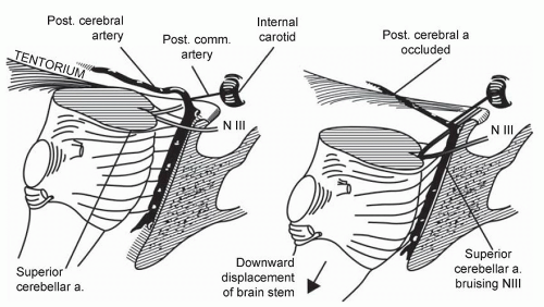
Figure 9.4 A schematic representation further depicting the close application of the tentorium and, therefore, the temporal lobe to the midbrain, the oculomotor nerve and the brainstem circulation. Source: ACS. ATLS Student Manual. Chicago: ACS, 1997.


Figure 9.5 A schematic representation of the sensory nerves of the brainstem with the midbrain devoid of major sensory nerves, the pons with the fifth nerve, important for the corneal reflex, and the eighth nerve at the pontomedullary junction, important for the oculocaloric reflexes. Source: Gray’s Anatomy, 28th edn. Baltimore: Lea & Febinger, 1968.

Direct measurement of brain tissue pO2 (PbtO2) is also under investigation as a cerebral metabolism monitor. Normal brain pO2 is in the range of 37 mm Hg, with severe hypoxia identified by values of 8 mm Hg or less. PbtO2 may be influenced by regional as well as global reductions in CBF (4).


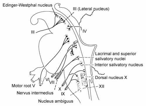
Figure 9.6 A schematic representation of the motor nerves of the brainstem, with the midbrain contributing the Edinger-Westphal nucleus for parasympathetic innervation of the pupil, and the third nerve that is important for oculovestibular and oculocaloric reflexes. The pons contributes the sixth nerve, also important for oculovestibular and oculocaloric reflexes, as well as the seventh nerve that is important for the corneal reflex. Source: Gray’s Anatomy, 28th edn. Baltimore: Lea & Febinger, 1968.


Table 9.2 Determinants of Increased Intracranial Pressure


  1. Increased CSF


    • Obstructed flow—occlusion of third ventricle


    • Obstructed reabsorption—occluded arachnoid granulations


  2. Increased brain cell volume


    • Trauma—contusion


    • Hyponatremia


    • Hypoxia—hypoperfusion


    • Infection—Reye syndrome


    • Mass lesions


    • Trauma—hemorrhage


      – Epidural


      – Subdural


      – Intraparenchymal


    • Tumors


  3. Increased cerebral blood volume


    • Increased CBF


    • Intracerebral vasodilation


ICP (normally <15 mm Hg) is determined by the following variables: cerebrospinal fluid (CSF) volume, cerebral blood volume (CBV), and brain cell volume (Table 9.2). The measurement of ICP and use of CPP as well as oxygen-related monitors to manage severe head trauma are discussed in the section on “Head Trauma”.


THE EFFECTS OF HYPOPERFUSION ON BRAIN FUNCTION

Depending on the magnitude and duration, decreased oxygen delivery to brain tissue can cause alterations in CNS function, ranging from agitation to brain death. The sudden cessation of cerebral circulation results in coma in 6-7 seconds, with the cerebral cortex suffering from lack of oxygen before the brainstem. Therefore, the initial state of coma is cortical coma.
The exact duration of absent circulation that will cause irreversible cortical and subsequent brainstem death is controversial.


Table 9.3 Glasgow Coma Scale


Physical Examination


Points


1. Eye-opening response


• Spontaneous


4


• To speech


3


• To pain


2


• None


1


2. Verbal response


• Oriented


5


• Confused, still answers


4


• Inappropriate words


3


• Incomprehensible sounds


2


• None


1


3. Best motor response


• Obeys


6


• Localizes


5


• Withdraws


4


• Abnormal flexion


3


• Abnormal extension


2


• None


1


After TBI, cerebral ischemia is a very early event that usually abates spontaneously or after evacuation of a mass lesion. Acute subdural hematomas and diffuse cerebral edema are at a greater risk for continued cerebral ischemia. The prognosis of patients with persistent ischemia is poor (2).

The relationship between Glasgow Coma Scale (GCS) (Table 9.3) and the CMRO2 in severe head trauma patients can be broadly characterized as follows (GCS <8 = coma) (5):


CMRO2 (normal 3.2)


GCS (normal 15)


1.1


3-4


1.5


5-6


1.5


7-8


Since CMRO2 is primarily determined by CBF, the association of a marked decrease in brain oxygen metabolism with severe neurologic malfunction following trauma suggests that decreased CBF aggravates brain cell dysfunction after trauma.


EFFECTS OF INFLAMMATION ON BRAIN FUNCTION

Inflammation-associated alterations in brain function are linked to both systemic and local inflammatory processes. For instance, systemic inflammation has been associated with decreased CBF, impairment of subcortical and cortical sensory-evoked potential pathways, release of brain injury biomarkers (S-100β and neuron-specific enolase), and decreased brain mitochondrial function (6, 7, 8, 9). Encephalopathy during severe systemic inflammation is common and correlated with increased mortality risk (8, 10).

TBI is characterized by rupture of the blood brain barrier (BBB) and the leakage of serum components and blood cells into the cerebral tissue that stimulate inflammatory cell migration and activation. These, in turn, engage endogenous microglia, astrocytes, and neurons in cell-to-cell interactions that augment the inflammatory state and the threat to cellular function and viability.


Table 9.4 Etiologies of Coma


  1. Cortical


    1. Metabolic


      • Hypoglycemia


      • Hypoxia


      • Hypercapnia


      • Hyperglycemia


      • Hyponatremia


      • Hypercalcemia


      • Narcotics


      • Barbiturates


      • Diazepam


      • Severe inflammation


      • Liver failure


      • Renal failure


    2. Infectious


      • Meningitis


      • Encephalitis


    3. Traumatic


      • Diffuse contusion


      • Subarachnoid bleed


  2. Brainstem


    1. Metabolic


      • Severe hypoxia—brainstem infarct


      • Severe drug overdose


    2. Traumatic


      • Supratentorial mass


      • Infratentorial mass


      • Direct injury


Activation of inflammatory mediators [especially Interleukin (IL)-6] has been linked to the extent of cerebral damage, BBB malfunction, leukocyte infiltration, and neurological outcome. However, too vigorous suppression of brain injury inflammation can also have detrimental effects on wound healing, cell regeneration, and recovery (11). In this respect, the brain, as with other tissues subjected to tissue injury and inflammation, can suffer from both too much and too little inflammatory response.


ETIOLOGIES OF COMA

Coma is categorized broadly into cortical and brainstem etiologies (Table 9.4). As mentioned previously, the brainstem is much more resistant to metabolic derangements and requires severe metabolic insults (i.e., cardiopulmonary arrest) to malfunction. Most brainstem dysfunction is secondary to nearby or direct traumatic injury or decreased basilar artery blood flow.

Broadly speaking, if physical examination is consistent with an intact brainstem (e.g., normal pupils, normal corneal reflexes, normal extraocular movements), the patient most likely has a cortical and, therefore, a metabolic coma (diffuse cortical trauma may cause similar findings). On the other hand, if any brainstem reflex is abnormal (e.g., one pupil larger and less reactive than the other, diminished corneal reflexes, absence of an extraocular movement), the patient is more likely to have a mass lesion pushing on the brainstem, or direct brainstem injury.


PHYSICAL EXAMINATION OF THE PATIENT WITH ALTERED BRAIN FUNCTION

As with all conditions, physical examination of the patient with neurological malfunction begins with vital signs. Vital signs may also alert the clinician to the presence of underlying neurological injury. Most critically ill patients are tachycardic. Bradycardia is unusual, especially
in trauma patients, and may indicate increased ICP or injury to the sympathetic nervous system in the high spinal cord. Similarly, hypertension is less common than hypotension, and can result from increased ICP. Since many critically ill surgical patients are assisted by a ventilator, respiratory status is a less useful indicator of neurological function, except for the broad categories of present or absent.


Table 9.5 Outline of Physical Examination for Brain Status


  • Vital signs


  • Glasgow Coma Scale


  • Pupil reflexes (constriction cranial nerve 3, dilation—entire brain stem)


  • Corneal reflexes (cranial nerves 5, 7)


  • Oculocaloric or oculovestibular reflexes (cranial nerves 3, 6, 8)


  • Posturing response


  • Gross motor, upper and lower extremity


  • Gross sensory, upper and lower extremity


  • Deep tendon and Babinski reflexes


Physical examination of brain neurological function begins with a global assessment of consciousness. The GCS (Table 9.3) is often used for this purpose, but it does not well describe the evaluation of the barely responsive or unresponsive patient, nor explains the localization of abnormalities. For such circumstances, eliciting brainstem and posturing reflexes can be a valuable adjunct (Table 9.5).

The pupillary reflex depends upon intact sympathetic (dilation from noxious stimuli) and parasympathetic (constriction from light) innervation (Fig. 9.7). The sympathetic nervous system arises in the hypothalamus, above the brainstem, runs through the brainstem, exits from the thoracic spinal cord, and follows the arterial supply to the eye. Constriction of the pupil is secondary to parasympathetic innervation that arises in the midbrain and accompanies the oculomotor nerve to the eye. Corneal reflexes represent sensation by the fifth and motor response by the seventh nerve, both in the pons. Oculovestibular and oculocaloric reflexes (Fig. 9.8) result from stimulation of the vestibular apparatus innervated by the eighth nerve at the ponto-medullary junction with response by the sixth and third nerves connected to the eighth nerve via the medial longitudinal fasciculus. Decorticate rigidity (arms point toward the cortex; Fig. 9.9) to noxious stimuli represents loss of cortical-spinal innervation either from cortical or internal capsule malfunction. Decerebrate rigidity (arms point away from the cortex; Fig. 9.10) usually represents at least partial, bilateral separation of midbrain function from higher centers (1).

Precise documentation of these aspects of the brain neurological examination is mandatory. Potentially confusing terminology such as “doll’s eyes” should be avoided. More useful is the statement “Oculovestibular reflex: medial and lateral rectus function bilaterally,” which describes precisely the test and the results.

Examination for lateralizing signs (i.e., one side moves differently from the other) is as important for identifying the risk for an intracranial mass lesion as is the recognition of an abnormal brainstem reflex (12).


CNS MALFUNCTION


Head Trauma

Head trauma, especially with coma, requires careful attention to the Airway-Breathing-Circulation (ABC) of resuscitation, as well as an orderly diagnostic approach. This may be difficult when confronted with other major injuries and the hemorrhage and unsightly appearance associated with head injury. Table 9.6 outlines the basic initial clinical approach to the patient with head injury, which may begin at the scene of injury and continue into the intensive care unit (ICU). The injured brain is particularly susceptible to further insults that decrease CBF or increase the local inflammatory reaction. In addition, the brain does not tolerate hypoxia secondary to inadequate pulmonary function. Therefore, rapid and aggressive attention to the
ABC of trauma and continuing attention to oxygenation and resuscitation of the circulation are as, if not more, important with brain injury as with any other injured tissues. Previous ideas that brain injury could be limited by providing minimal resuscitation of the circulation are not supported by epidemiologic studies that demonstrate an increased risk of death and neurologic disability in brain-injured patients who suffer from hypotension anytime during the early post-injury period (13, 14, 15, 16).

Only gold members can continue reading. Log In or Register to continue

Stay updated, free articles. Join our Telegram channel

Jul 5, 2016 | Posted by in CRITICAL CARE | Comments Off on The Nervous System

Full access? Get Clinical Tree

Get Clinical Tree app for offline access