CHAPTER 74 TECHNIQUES IN THE MANAGEMENT OF COMPLEX MUSCULOSKELETAL INJURY: ROLES OF MUSCLE, MUSCULOCUTANEOUS, AND FASCIOCUTANEOUS FLAPS
Injuries involving skin and subcutaneous tissue loss require reconstructive solutions. In many cases, skin grafting alone may be sufficient. However, when skeletal fractures, tendons, viscera, or hardware is exposed, vascularized soft tissue coverage of the wound using muscle, musculocutaneous, and fasciocutaneous flaps is the preferred technique. The objective is to provide wound healing, optimal function, and the best possible aesthetic result.
DIAGNOSIS
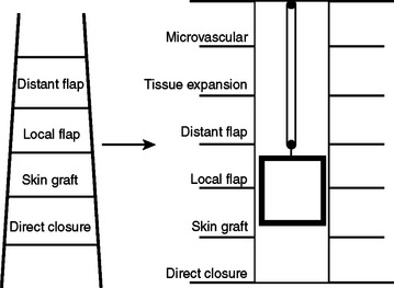
The concept of the “reconstructive ladder” recommends the use of the least invasive repair that will satisfactorily close a wound. The methods begin with secondary healing and progress through skin grafting to microvascular free-tissue transfer. This paradigm has been largely supplanted by the “reconstructive elevator” concept.1 With this approach, the surgeon evaluates the wound and decides on the best option, not necessarily the least complicated one (Figure 1).
ANATOMY
The most important anatomical aspect of muscle flaps is their vascularity. Because blood supply is usually the limiting factor in flap success, flaps are most often categorized by the vascular system on which they are based. McGregor proposed the concept of “random” and “axial” pattern flaps based on the importance of the presence or absence of a major vessel running along the axis of the flaps.2 Random pattern flaps do not incorporate a dominant vascular supply, relying on the networks of small-diameter vessels to sustain the transferred tissue. They are limited in size and may require delay for successful transfer. Axial pattern flaps incorporate an anatomically recognized arteriovenous system running along the long axis of the tissue which permits successful transfer of vascularized flaps with high length-to-breadth ratios. They obtain their vascular supply from the musculocutaneous and fasciocutaneous systems, both of which rely on multiple “perforator” arteries. Knowledge of muscle vascular anatomy is helpful in predicting the viability of overlying skin territories based on such perforating vessels.
The now classic schema of Nahai and Mathes has divided muscles into groups according to their principal means of blood supply.2 A type I muscle, such as the gastrocnemius or tensor fascia lata (TFL), is supplied by a single pedicle. A type II muscle, such as the trapezius or gracilis, has a dominant pedicle, with one or more minor pedicles. A type III, the serratus anterior (SA) or gluteus maximus (GM), for example, has dual dominant pedicles. A type IV, such as the tibialis anterior (TA) or sartorius, has segmental pedicles. The type V, such as the internal oblique muscle or latissimus dorsi (LD), has a dominant pedicle, with secondary segmental pedicles. Most muscles fall into the type II group. Types I, III, and V are the most reliable because complete muscle viability can be sustained by a single vessel. Sometimes the muscle territory of a minor pedicle is poorly captured by the dominant pedicle in a type II muscle. Owing to their segmental means of perfusion, type IV muscles would potentially only allow small flaps that have limited application.
SURGICAL MANAGEMENT: PRIMARY FLAPS
Musculoskeletal trauma will be grouped into five soft tissue coverage regions: head and neck, upper extremity, chest and trunk, abdomen, and the lower extremity (Table 1).
Head and Neck
The pectoralis myocutaneous flap can be designed with a skin paddle centered over the lower portion of the muscle. It can be used to resurface the neck, cheek, oral cavity, palate, tonsillar area, and nasopharynx, tongue, floor of mouth, mandible, and cervical area. The flap has an upward arc of rotation of 180 degrees and may be raised as high as the orbits; however, in practicality, it is difficult to secure the closure without a significant downward pull on the muscle. The flap can be modified with an extended random skin component or with two separate skin paddles that can be divided. A rib may be harvested with the flap for bony reconstruction. Higher elevation of the flap can be performed with the division of the clavicle. The pectoralis was one of the early workhorse flaps, but it has largely been supplanted by free flaps.
Stay updated, free articles. Join our Telegram channel

Full access? Get Clinical Tree


