0.12 seconds.
- A wide complex rhythm suggests an aberrancy in the normal conduction system and is usually the result of one of the following:
- A preexisting or rate-related abnormality within the normal conduction system (e.g., bundle branch block).
- An accessory pathway (e.g., Wolff–Parkinson–White syndrome [WPW]).
- A preexisting or rate-related abnormality within the normal conduction system (e.g., bundle branch block).
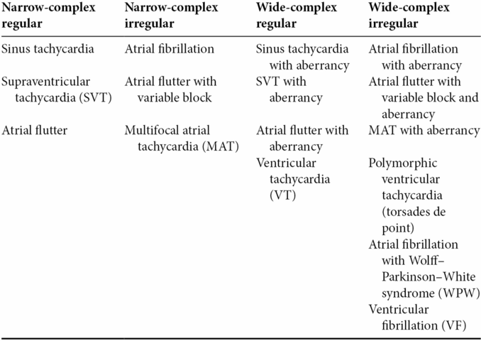
Table 21.1. Classification of common tachyarrhythmias
Presentation
Classic presentation
- Ironically, the “classic presentation” mostly consists of nonspecific symptoms. Patients may complain of palpitations, chest pain, lightheadedness, dyspnea, or nonspecific weakness.
- Further evaluation will reveal a rapid heart rate on physical examination or on the electrocardiogram (ECG).
Critical presentation
- Patients with unstable tachyarrhythmias present with signs and symptoms of hypoperfusion and hemodynamic compromise while still maintaining a palpable pulse:
- Hypotension
- Altered mentation
- Ischemic chest pain
- Pulmonary edema.
- Hypotension
- Patients who do not have a palpable pulse are deemed to be in cardiac arrest and are treated according to Advanced Cardiovascular Life Support (ACLS) guidelines.
Diagnosis and evaluation
- Primary evaluation consists of performing an ECG with a rhythm strip.
- Once tachyarrhythmia is confirmed, consideration should be given to whether the arrhythmia has an underlying noncardiac etiology such as a toxic ingestion or a metabolic disturbance.
- Clinical history is useful and important. Attention should be paid to the patient’s medical history and potential use of QT-prolonging medications.
- A chest radiograph and basic blood work may be helpful in identifying a metabolic or infectious etiology of the arrhythmia.
- Clinical history is useful and important. Attention should be paid to the patient’s medical history and potential use of QT-prolonging medications.
- The type of arrhythmia can be determined on the basis of the ECG:
- Sinus tachycardia:
- Narrow-complex tachycardia.
- Regular rate greater than 100 bpm.
- Rates >160 bpm are not typically attributable to sinus tachycardia.
- Each P wave is associated with a QRS complex.
- There is a fixed P-R interval.
- Narrow-complex tachycardia.
- Sinus tachycardia:
- Supraventricular tachycardia (SVT):
- Narrow-complex tachycardia.
- Regular rate between 140 and 250 bpm.
- P waves may be present but difficult to see due to the rate.
- The most common cause of SVT is atrioventricular nodal reentrant tachycardia (AVNRT).
- SVT may also present as a wide-complex tachycardia if it is associated with a rate-related or preexisting bundle branch block. This is often referred to as “SVT with aberrant conduction” and may mimic VT.
- Narrow-complex tachycardia.
- Atrial fibrillation:
- Atrial fibrillation is the most common cardiac arrhythmia.
- Usually narrow complex, but can present with a wide QRS in the presence of underlying disease of the conduction system.
- Irregularly irregular rhythm with absent P waves and atrial rates varying from 400 to 700 bpm.
- Ventricular response is usually 120–180 bpm.
- Irregularly irregular rate distinguishes AF from other arrhythmias, even when the complex is wide.
- Atrial fibrillation is the most common cardiac arrhythmia.
- Atrial flutter:
- On a spectrum of sinus node dysfunction with atrial fibrillation.
- Usually narrow complex.
- Presents with a classic “sawtooth” pattern of P waves.
- The atrial rate ranges between 250 and 350 bpm.
- Atrial flutter is associated with varying degrees of AV block and can present as a regular or regularly irregular rhythm.
- Most often, the atrial rate is regular at 300 bpm with a 2:1 block, producing a regular ventricular rate of 150 bpm.
- On a spectrum of sinus node dysfunction with atrial fibrillation.
- Multifocal atrial tachycardia:
- Irregularly irregular tachyarrhythmia.
- Diagnosed by the presence of at least three different P wave morphologies with varying P-R intervals.
- MAT is almost always seen in the elderly and those with pulmonary disease. It is also associated with hypomagnesemia, hypokalemia, and coronary artery disease.
- Irregularly irregular tachyarrhythmia.
- Ventricular tachycardia:
- Wide complex regular tachyarrhythmia.
- Ventricular rate is greater than 120 bpm.
- VT can be monomorphic or polymorphic:
- Monomorphic VT usually presents with rates between 120 and 300 bpm.
- Polymorphic VT usually has rates >200 bpm.
- Torsades de pointes is a polymorphic VT with a prolonged QT. On the ECG, the QRS complex appears to be twisting around an axis. It is a subtype, not a synonym, of polymorphic VT.
- Monomorphic VT usually presents with rates between 120 and 300 bpm.
- Wide complex regular tachyarrhythmia.
- All wide-complex regular tachycardias are potentially life threatening and should be considered VT until proven otherwise.
- Ventricular fibrillation:
- Wide complex irregular tachyarrhythmia.
- Always associated with unstable or pulseless patient.
- Wide complex irregular tachyarrhythmia.
Critical management
- In patients with tachyarrhythmias, critical management actions include
- Assessment of overall stability with ABCs
- ECG and continuous telemetry
- Intravenous access
- Placement of pacer/defibrillation pads on the patient in anticipation of potential deterioration
- Assessment of overall stability with ABCs
- Definitive therapy will vary depending on the underlying rhythm.
- Commonly used medications as well as their dosage are presented in Table 21.2.
- Sinus tachycardia
- The primary goal with a patient in sinus tachycardia (ST) is to treat the underlying condition rather than the tachycardia itself.
- The primary indication for rate control in ST is during acute myocardial infarction, where tachycardia is associated with worse outcomes. Nodal agents, particularly beta-blockers, are useful in this setting.
- The primary goal with a patient in sinus tachycardia (ST) is to treat the underlying condition rather than the tachycardia itself.
- Paroxysmal supraventricular tachycardia
- Generally unrelated to an underlying cause.
- Rhythm control is the primary intervention.
- Vagal maneuvers can be attempted prior to pharmacological therapy.
- Adenosine is the medication of choice as it is both diagnostic and therapeutic.
- Adenosine is metabolized quickly by nonspecific esterases in the plasma and therefore should be pushed quickly via the intravenous access closest to the heart.
- Generally unrelated to an underlying cause.
- If adenosine fails, consider synchronized cardioversion or rate control with agents that slow conduction through the AV node.
- Atrial fibrillation and atrial flutter
- These rhythms are modulated by sinus automaticity and, like sinus tachycardia, can be driven by underlying etiologies.
- Rate control is the primary treatment modality
- Nodal blockers and digoxin are reasonable options.
- Amiodarone is an appropriate alternative.
- Of note, rapid atrial fibrillation with a wide QRS complex suggestive of WPW should not be treated with AV nodal blocking agents. In this setting, procainamide or synchronized cardioversion should be used.
- Nodal blockers and digoxin are reasonable options.
- These rhythms are modulated by sinus automaticity and, like sinus tachycardia, can be driven by underlying etiologies.
- Sinus tachycardia
- Multifocal atrial tachycardia
- Initial therapy for MAT should be aimed at treating the underlying cause such as hypomagnesemia, hypokalemia, pulmonary, or cardiac disease.
- Pharmacological therapy is indicated if the arrhythmia is causing significant symptoms such as ischemia, hypoxia, heart failure, or shock.
- Nodal blockers can be used, though their efficacy is limited.
- Initial therapy for MAT should be aimed at treating the underlying cause such as hypomagnesemia, hypokalemia, pulmonary, or cardiac disease.
- Ventricular tachycardia
- In stable patients with monomorphic VT, procainamide or amiodarone can be used.
- Polymorphic VT is often caused by myocardial ischemia. Diagnosis and treatment of potential myocardial infarction should be pursued.
- Treatment of torsades de pointes is aimed at decreasing the QT interval.
- Intravenous magnesium sulfate is the first-line treatment.
- Overdrive pacing also shortens the QT interval and can be used if magnesium therapy is ineffective. It can be achieved by transcutaneous pacing or with pharmacological agents such as isoproterenol.
- Intravenous magnesium sulfate is the first-line treatment.
- Ventricular fibrillation:
- VF is an unstable rhythm that should be primarily managed by defibrillation.
- ACLS should be initiated promptly in all patients exhibiting this rhythm.
- VF is an unstable rhythm that should be primarily managed by defibrillation.
- In stable patients with monomorphic VT, procainamide or amiodarone can be used.
Table 21.2. Common medications for the treatment of tachyarrhythmias
Related posts:


Stay updated, free articles. Join our Telegram channel

Full access? Get Clinical Tree


