SURGICAL ANATOMY OF THE ABDOMEN AND RETROPERITONEUM

CHAPTER 44 SURGICAL ANATOMY OF THE ABDOMEN AND RETROPERITONEUM


The abdominal cavity and the retroperitoneum lie immediately adjacent to one another. Some organs such as the small bowel and the colon have portions within the abdominal cavity while other parts are within the retroperitoneum. Vascular structures such as the superior mesenteric artery and vein course through both body compartments as well. A thorough knowledge of the anatomy of both the abdomen and retroperitoneum is critical for a rational operative approach to torso injuries.


Injuries to both the abdomen and retroperitoneum are generally approached via the same incision. Some structures such as the supraceliac aorta or the pelvic vasculature may require a counter incision such as a thoracotomy, a groin exploration, or a direct retroperitoneal incision to identify and repair specific injuries. In this chapter, we review the pertinent anatomic considerations of both the abdomen and retroperitoneum. In particular, we will stress functional anatomic considerations that are important during operative trauma surgery.


MAKING THE INCISION


The abdomen and retroperitoneum are generally explored via a generous midline incision. The incision should be made from the xiphoid to pubis, unless a specific already-diagnosed injury is to be treated. This gives the greatest access to all of the structures in the abdomen and retroperitoneum. Several additional options exist in order to reach specific areas. A thoracoabdominal approach gives access to certain structures high in the abdomen. This generally involves a seventh or eighth interspace anterolateral thoracotomy that is brought down to the sternum. The ribs are divided flush with the sternum. The diaphragm is then taken down off the chest wall radially. Approximately one to two inches should be left on the chest wall for diaphragmatic reconstruction later. On the left side, the diaphragm should be taken down all the way to the aorta. A left-sided thoracoabdominal approach is probably the best exposure for the supraceliac aorta. A right-sided thoracoabdominal incision increases the exposure of the posterior portion of the right lobe of the liver. Exposure of the retrohepatic cava is also enhanced with the use of a right-sided thoracoabdominal approach.


Occasionally, the midline incision is extended up into a medium sternotomy. This gives access to the anterior mediastinal structures. If an atriocaval shunt is to be used to treat a retrohepatic caval injury, a sternotomy is probably the best approach. In addition, if one wishes to control the inferior vena cava within the pericardium to achieve complete vascular isolation of the liver, a sternotomy or a right-sided thoracoabdominal incision will give the surgeon adequate access to perform that maneuver.


Exposure of the deep pelvic vasculature can also be difficult through a standard laparotomy. Several options exist to increase that exposure. A groin incision allows for vascular control of the common femoral artery and vein at the level of the inguinal ligaments. A combination of a full laparotomy and groin incision can aid in repair of a vascular injury immediately adjacent to the inguinal ligaments. Another option is to perform a retroperitoneal incision similar to those used for renal transplant. This hockey stick incision comes down through the retroperitoneum and exposes the distal iliac artery and vein. Distal pelvic vascular repair can then be accomplished via the combined incisions. While rare, if a transplant incision is combined with a midline incision, the bridge of skin, subcutaneous tissue, and fascia between the two incisions can become ischemic and infarct.


EXPLORING THE ABDOMEN


As the fascia is divided at the time of laparotomy, it is helpful to look at the peritoneum. If it is bulging, and has a bluish discoloration, that generally means there is a tense hemoperitoneum that will be released as the peritoneum is opened. Blood should be aspirated into a cell saver circuit. The most commonly injured organs after blunt trauma are the liver and spleen. They can be quickly assessed for injury by palpating the right upper quadrant and left upper quadrant. The surgeon can then sweep the small bowel and colon medially, which identifies any large retroperitoneal hemorrhage. If the base of the small bowel mesentery is examined, any mesenteric injury can be identified.


It is imperative to fully mobilize the spleen in order to be able to fully inspect all surfaces. This allows for intelligent decision making as to whether splenectomy or splenorrhaphy will be wise (Figure 1). All of the ligamentous attachments must be divided in order to get the spleen up to the anterior abdominal wall.


A similar strategy is important when inspecting the injured liver. The falciform ligament should be taken down all the way to the level of the vena cava. Both triangular ligaments must be completely incised. This leaves the liver suspended only on the hepatic veins and the portal structures. This allows the surgeon to rotate the liver up out of the deep recesses of the right upper quadrant, which is especially important when evaluating injuries to the posterior right lobe. In addition, one should evaluate whether sternotomy and/or right thoracotomy with takedown of the diaphragm will be needed for added exposure.


Most blunt liver injuries are now treated nonoperatively. Bleeding from higher-grade liver injury is often persistent and of significant volume. In these cases, manual compression by direct pressure or packing will often temporize the bleeding. The Pringle maneuver is often diagnostic, and it achieves temporary hemostasis. The Pringle maneuver involves placing a non-crushing vascular clamp across the hepaticoduodenal ligament, thereby occluding both portal venous and hepatic arterial inflow. Once the clamp is applied, the liver can be reinspected and repair undertaken under better circumstances. If bleeding persists despite portal compression, the surgeon must suspect a retrohepatic inferior vena cava injury, hepatic vein injury, or anomalous hepatic arterial anatomy (10%ā€“25%). The main or right hepatic artery comes off the superior mesenteric artery (SMA) in 10%ā€“20% of cases, as the accessory right hepatic artery comes off the SMA in about 5% of cases. In addition, an anomalous left hepatic artery comes off the left gastric artery in nearly 5% of cases.


While often quite effective, the Pringle maneuver has some drawbacks. The Pringle maneuver produces global ischemia of the liver, potentially worsening hepatic function in the postoperative period. While there is some evidence that the liver can withstand several hours of warm ischemia during elective hepatic resection, the same may not be true for the patient in shock. The Pringle maneuver can also be cumbersome, limiting exposure. In addition, in severe liver injuries there almost always is a component of hepatic venous injury limiting the utility of the Pringle maneuver.


In more severe cases of liver injury, one may also consider the Heaney maneuver, which involves complete vascular isolation of the liver. In this technique, vascular clamps are placed on the cava, above and below the liver. When combined with the Pringle maneuver and/or supraceliac aorta clamping, this should provide for a relatively dry field in order to attempt to definitively repair the liver, and the hepatic venous injury. While this can be effective, it may be difficult to dissect out the suprahepatic vena cava in order to place a clamp, unless the diaphragm has been divided or the pericardium opened via a thoracotomy or sternotomy. The infrahepatic cava must be occluded distal to the junction of the renal vein. If the clamp is placed too low, flow from the renal vein into the cava will perpetuate bleeding and continue to make exposure extremely difficult. Occasionally, complete vascular isolation can be combined with a venous bypass circuit to allow for venous return to the heart (Figure 2).
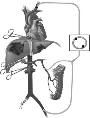


Perhaps the greatest downside to the use of complete vascular isolation is the rapid and profound decrease in venous return to the heart. In these critically ill patients, this can occasionally produce cardiovascular collapse and/or cardiac arrest. If the technique is to be used, all resuscitation lines must be placed in the upper extremity or the mediastinal veins.


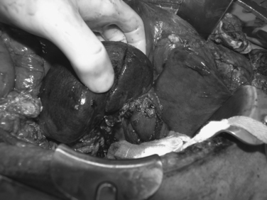
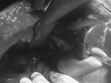
An intermediate solution to injuries with a hepatic vein component is manual compression medial to the injury. This should control all inflow to the injured segment. The injured liver can then be debrided. The hepatic vein can then be identified and ligated when pressure is relaxed. The more central hepatic arterial and portal venous branches can be ligated as well using the same temporary relaxation of hand held pressure (Figure 3).


The anterior portions of the duodenum and the head of the pancreas are in the abdomen. The posterior portion of the duodenum and the head of the pancreas are in the retroperitoneum. A full Kocher maneuver (incising the lateral peritoneal reflection and completely mobilizing the duodenum and pancreas) is necessary to evaluate both structures for injuries. The body of the pancreas lies within the lesser sac. It is necessary to widely open the lesser sac to examine the posterior stomach, as well as the anterior aspect of the body of the pancreas. This is best accomplished by dividing the gastrocolic omentum.
The gastrocolic omentum can be divided all the way up the greater curvature of the stomach to the level of the gastroesophageal (GE) junction. This requires taking the short gastric vessels adjacent to the spleen. When the gastrocolic omentum has been completely divided, the surgeon has good access to the lesser sac, allowing inspection of the posterior wall of the stomach. It is necessary to carefully inspect the stomach, particularly around the area of the GE junction in order to avoid missing small injuries. If necessary, the intra-abdominal portion of the esophagus can be mobilized on a Penrose drain to aid in exposure. We attempt to triangulate the stomach using sponge sticks to flatten out both the anterior and posterior aspect of the stomach adjacent to the GE junction. This allows for complete inspection and avoids missing a subtle injury high up on the stomach.


The area of the porta contains the hepatic artery, portal vein, and common bile duct. The portal structures are covered with peritoneum. When the peritoneum is opened, the common bile duct is generally the first structure encountered. The hepatic artery can be identified by its palpable pulse and the thrill usually present within it. The portal vein lies posterior to the common bile duct. Each of these can be individually isolated and examined for injury.


It is necessary to completely evaluate the small intestine to avoid missing an injury. Complete evaluation of the bowel involves running the small bowel using a hand over hand technique. The bowel should be flipped with each inspection to be sure both sides have been completely evaluated. Spreading the bowel out allows for inspection of the corresponding mesentery. Once major vascular injuries and solid visceral injuries are controlled, the small bowel should be examined next. We generally re-examine the small bowel before closing to avoid missing an injury.

< div class='tao-gold-member'>

Stay updated, free articles. Join our Telegram channel

Jul 7, 2016 | Posted by in CRITICAL CARE | Comments Off on SURGICAL ANATOMY OF THE ABDOMEN AND RETROPERITONEUM

Full access? Get Clinical Tree

Get Clinical Tree app for offline access