CHAPTER 17 Suprascapular block
Surface anatomy
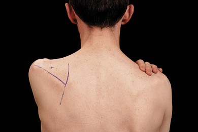
The spine of the scapula is the main bony landmark for the suprascapular block (Fig. 17.1). It can be palpated from the medial aspect of the scapula and followed laterally and superiorly to the acromion. Two centimeters superior to the midpoint of the spine of the scapula is the needle insertion point.
Sonoanatomy
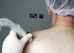
An initial scan is performed in the sagittal orientation at the superior medial border of the scapula to identify the pleura. Scanning proceeds laterally with this transducer orientation. Where the scapula moves beyond the lung field is noted. The ultrasound transducer is now placed parallel to the scapular spine (Fig. 17.2) such that the scapular spine is visualized as a superficial hyperechoic line (Fig. 17.3). By moving the transducer cephalad the suprascapular fossa is identified (Fig. 17.4). While imaging the supraspinatus muscle and the bony fossa underneath, the ultrasound transducer is slowly moved laterally (maintaining a transverse transducer orientation) to locate the suprascapular notch (Fig. 17.5). The suprascapular nerve is seen as a round hyperechoic structure at 4–6 cm depth beneath the transverse scapular ligament in the scapular notch (Fig. 17.5).
< div class='tao-gold-member'>
Related posts:


Stay updated, free articles. Join our Telegram channel

Full access? Get Clinical Tree


