SOFT TISSUE INFECTIONS

CHAPTER 80 SOFT TISSUE INFECTIONS


Soft tissue infections occur frequently and account for approximately 48.3 in 1000 outpatient visits. The severity of these infections varies from trivial to life-threatening.


Severe soft tissue infections have been described throughout the medical literature since ancient times. Necrotizing fasciitis was described in the fifth century BC by Hippocrates, though Wilson coined the term in 1952.


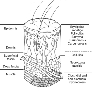
Much of our knowledge regarding the treatment of soft tissue infection has been based on the experience gained during military conflicts. For instance, hospital gangrene was described first by Joseph Jones, a Confederate surgeon during the Civil War. The treatment of battlefield infections has influenced civilian practice. This review will begin with a description of the anatomically more superficial infections and progress to the deeper, life-threatening infections (Figure 1).


SUPERFICIAL INFECTIONS


Superficial infections are limited anatomically to the epidermis and the dermis. These infections can occur spontaneously or secondary to minor trauma. Impetigo usually presents with vesicles that leak, producing a thick yellow crust. These lesions are typically located on the face, neck, and extremities. Staphylococcus aureus and Streptococcus pyogenes are the most common causative agents. Bullous impetigo, like impetigo, is most commonly caused by S. aureus. It is characterized by small vesicles that coalesce to form large bullae.


Folliculitis develops as an infection of the hair follicle. The lesions appear as pustules or papules, commonly on the extremities, scalp, or beard. Whirlpool folliculitis is associated with immersion in inadequately chlorinated pools, whirlpools, or hot tubs. Diffuse pustules are seen. Pseudomonas aeruginosa is the classic causative agent. Swimmer’s itch is a folliculitis that develops after freshwater exposure.


Furuncles, deeper inflammatory nodules, can develop from folliculitis. S. aureus is often the causative agent. Carbuncles are coalescing furuncles formed by connecting sinuses. The nape of the neck is the most common anatomic location. S. aureus is most often isolated. Patients often have comorbidities such as diabetes mellitus, alcoholism, immunosuppression, or malnutrition. Systemic infection may result from these lesions.


Cellulitis is an inflammation of the subcutaneous tissue. There is erythema, pain, and edema of varying severity. A portal of entry for the bacteria is usually present. It may be as mundane as a crack in the skin from dryness or athlete’s foot. More substantial trauma may be involved, such as shotgun or shrapnel penetrations. Systemic symptoms may manifest and include fever, chills, malaise, and (infrequently) organ failure. Streptococcus (Groups A, B, C, and G) as well as staphylococcus are frequent culprits. Areas of compromised venous or lymphatic drainage are prone to this infection, and recurrence is common. Pelvic radiation or having had lymphadenectomy, mastectomy, or venectomy makes the development of cellulitis more likely.


DEEP INFECTION


Deeper infections are more frequently life- and limb-threatening. The literature concerning the potentially life-threatening infections is confusing. Multiple terms are used to describe the same disease, depending on the clinical setting in which it arises. The treatment is the same regardless of the term used to describe it. It therefore seems that anatomic classification is more logical and easily remembered. This review uses anatomic characterizations and relate them to older terminology as necessary.


In the medical literature, deep structure infection has masqueraded under a variety of pseudonyms. The term “necrotizing soft tissue infection” (NSTI)is used here. Table 1 lists a variety of terms that may appear in the medical literature to describe this infective process.


Table 1 Terms Used to Describe NSTI


Meleney’s synergistic gangrene Hospital gangrene
Streptococcal gangrene Fournier’s gangrene
Gas gangrene Acute dermal gangrene
Suppurativa fasciitis Necrotizing erysipelas
Phagedena Phagedena gangrenosum
“Flesh-eating disease” Necrotizing fasciitis
Clostridial cellulitis

All the terms describe an infection involving the subcutaneous fat and fascia, with variable skin involvement. The incidence of this infection is not known. A recent report quotes World Health Organization statistics of 500–1500 cases of necrotizing fasciitis annually. Though uncommon, it is not rare. The Centers for Disease Control has monitored Group A streptococcal infections and estimated 10–20 cases per 100,000 population. In Ontario, Canada, a population study estimated an incidence of 0.6 per 100,000.


CLINICAL PRESENTATION


Signs and symptoms of NSTI can be quite nonspecific (Table 2). Pain, erythema, and swelling of the affected area are most frequently present. This same constellation of symptoms may be seen in pathologic processes that have a much more benign course and respond effectively to antibiotic therapy alone. “Hard signs” of necrotizing infection include tense erythema, bullae, skin discoloration, and crepitus, pain out of proportion to examination, or anesthesia of the affected area. Unfortunately, many of these are late signs and indicate that the infective process is well established or they occur only in a small percentage of patients.


Signs of systemic toxicity may also be present. These may include pyrexia, tachycardia, hypotension, and organ dysfunction. The progression of symptoms may be rapid over the course of hours to days or more indolent over the course of days to weeks. The rate of progression of symptoms may be ameliorated by partial treatment. Some suggest classifying the disease by its clinical course. Fulminant disease presents in patients with acute onset and rapid progression over the course of hours with shock. Acute disease presents with large surface area involvement and over the course of days. Subacute disease presents for weeks and is usually localized. Differentiating this process from cellulitis or simple abscess can be a challenge. Clinically, failure to improve with appropriate antibiotics or worsening systemic toxicity portends this diagnosis (Table 3).


Table 3 Bacteriology


Rapid progression:


Slower progression:


< div class='tao-gold-member'>

Stay updated, free articles. Join our Telegram channel

Jul 7, 2016 | Posted by in CRITICAL CARE | Comments Off on SOFT TISSUE INFECTIONS

Full access? Get Clinical Tree

Get Clinical Tree app for offline access