| Type | Etiology |
|---|---|
| Mechanical | A physical barrier exists that blocks normal intestinal flow |
| Closed-loop | Two sequential sites of obstruction block a portion of bowel, usually through a hernia or adhesive band |
| Functional obstruction/adynamic ileus | Disturbance in gut motility, failure of peristalsis, most often caused by laparotomy, critical illness, or prolonged opiate use |
| Pseudo-obstruction | Disturbance of gut motility associated with an underlying condition, but without an underlying lesion. Examples: amyloidosis, hyperthyroidism, hypokalemia, etc. |
Table 43.2. Common causes of SBO (percent of all cases)
| Postoperative adhesions | 50% |
| Hernias | 15% |
| Neoplasms | 15% |
| Intussusception | 5% |
| Other | 15% |
Presentation
Classic presentation
- Crampy, paroxysmal abdominal pain with abdominal distension, vomiting, and inability to pass flatus in a patient with prior abdominal surgery, abdominal neoplasm, or hernia.
- Vomiting may only occur if the obstruction is proximal.
- Bowel dilatation causes increased secretory activity that causes accumulation of fluid in the proximal bowel and increased peristalsis. As a result, frequent loose stools and flatus may be seen in early bowel obstructions.
- Loss of flatus is a late finding.
Critical presentation
- Increased bowel distension causes increased bowel wall edema that results in significant fluid shifts. This may lead to:
- Hypovolemic shock
- Electrolyte abnormalities
- Severe acidosis.
- Hypovolemic shock
- Bowel perforation and peritonitis:
- Bowel perforation may be due to increased intraluminal pressure. In addition, if the arterial supply to the bowel is compromised (severe edema, physical obstruction of mesenteric pedicle), bowel wall ischemia and necrosis will follow.
- Severe septic shock due to bacterial translocation.
- Multi-organ system failure due to primary SIRS response or subsequent shock states.
- Age, treatment delay, and preexisting comorbidities are all associated with a worse prognosis.
Diagnosis and evaluation
- History
- A thorough history should be taken, with particular attention paid to prior SBOs, abdominal surgeries, hernias, cancer, and opiate use.
- Physical examination
- Vital signs: Fever, tachycardia, hypotension, tachypnea (compensate for metabolic acidosis or in response to pain).
- Abdominal examination:
- Visually inspect the abdomen for scars and distension.
- Auscultate for high-pitched bowel sounds.
- Assess for peritoneal findings and distension.
- Palpate and visually inspect for hernias.
- Visually inspect the abdomen for scars and distension.
- Rectal examination: Consider a rectal examination with evaluation for occult blood, although diagnostic yield may be low and classically the rectal vault will be empty.
- Vital signs: Fever, tachycardia, hypotension, tachypnea (compensate for metabolic acidosis or in response to pain).
- Laboratory evaluation
- Findings will not be specific to bowel obstruction. Results may show evidence of dehydration, acidosis, renal failure, leukocytosis, etc.
- Imaging
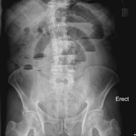
- Plain films (Figure 43.1)
- Plain radiography has poor sensitivity and specificity (46% sensitivity, 66% specificity).
- Indicated in almost all patients with clinically suspected SBO.
- Findings include multiple air–fluid levels, dilated loops of bowel, and absent colonic gas. (Figure 43.1)
- As good as CT scan for “high-grade” obstruction.
- Poor sensitivity for “low-grade” obstruction.
- Cannot differentiate between strangulation and simple obstruction.
- Plain radiography has poor sensitivity and specificity (46% sensitivity, 66% specificity).
- CT scan:
- CT has 80–90% sensitivity, 70–90% specificity.
- Can often diagnose obstruction when plain radiography is inconclusive.
- May be helpful to further characterize previously diagnosed obstruction.
- May show cause of obstruction, such as a mass or occult hernia.
- Findings not present on simple radiography may include bowel wall thickening, pneumatosis, portal venous gas.
- Allows determination of “transition point” which aids in surgical planning.
- May show alternative diagnoses.
- Does not necessarily need oral contrast as intraluminal fluid will serve this purpose.
- One must weigh the benefit of CT against potential risks of traveling for a sick patient and possible delay in surgical intervention.
- CT has 80–90% sensitivity, 70–90% specificity.
- Ultrasonography:
- US has 93% sensitivity, 100% specificity.
- With proper training, bedside ultrasound compares favorably with traditional plain radiography.
- Advantages include that it can be done rapidly at the bedside, it is less costly and less invasive, and there is no need for risky patient travel.
- Findings include
- Fluid-filled bowel loops >2.5 cm (90% sensitivity, 83% specificity)
- Decreased peristalsis (27% sensitivity, 97% specificity).
- Fluid-filled bowel loops >2.5 cm (90% sensitivity, 83% specificity)
- US has 93% sensitivity, 100% specificity.
- SBO that is confirmed with radiography or ultrasound in the setting of a good clinical presentation or with distinct clinical signs of obstruction or peritonitis will likely need operative management. Further imaging should not be necessary.
- Plain films (Figure 43.1)

Figure 43.1. Small bowel obstruction on plain film demonstrates multiple air–fluid levels in dilated loops of bowel with no air in the rectum. (Image by Mike Cadogan from the Global Medical Education Project [GMEP.org]. This image is protected under a Creative Commons CC BY-NC-SA 3.0 license.)
Stay updated, free articles. Join our Telegram channel

Full access? Get Clinical Tree


