Mechanical
Biological
Electrode fracture or migration for SCS
Infection
Catheter fracture or migration for ITDDS
Bleeding
Battery failure for SCS or ITDDS
Nerve injury
Programming or pump-fill errors for ITDDS
Seroma
Hygroma
30.3 Etiology and Pathogenesis
A seroma is a postoperative fluid collection resulting from an accumulation of serous fluid on the skin’s surface (Figs. 30.1 and 30.2). When it develops at the site of a surgical incision, it contributes to poor wound healing. The etiology of seroma formation is multifactorial; lymphatic disruptions, shearing between tissue surfaces, mediators of inflammation, and the creation of surgical dead space have been implicated [2]. Macrophages and polymorphonuclear leucocytes along with the release of histamines and prostaglandins result in vasodilatation of blood vessels and formation of interstitial fluid [2]. The fluid, known as serum, can be clear or yellowish in color.
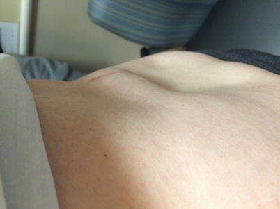
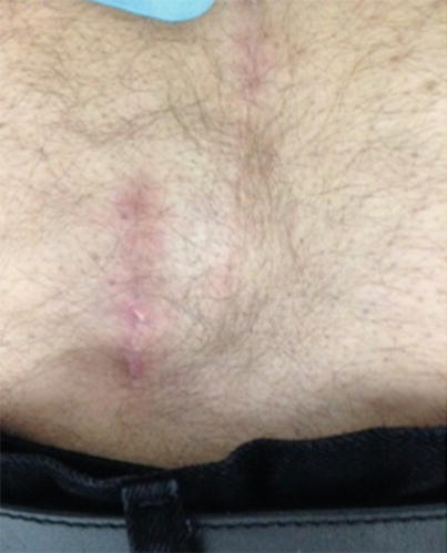
Fig. 30.1
Seroma formation along midline incision following implantation of an intrathecal drug delivery system. It developed around the incision used for placement of the intrathecal portion of the system. Image from Dr. Elmofty personal library
Fig. 30.2
Seroma formation along midline incision following implantation of an intrathecal drug delivery system. The patient denies positional headaches and placement of an abdominal binder has significantly reduced the size of the seroma. Image from Dr. Anitescu personal library
A hygroma is a subcutaneous collection of cerebrospinal fluid (CSF) . CSF leaks when the dura is injured, for example, from an introducer during placement of an intrathecal catheter. The needle is larger than the catheter inserted, thus creating an incomplete tissue seal around the intrathecal catheter. CSF can flow from the intrathecal space down the catheter and may collect anywhere along the catheter, including the pump pocket.
30.4 Risk Factors
Certain factors increase the risk for developing a seroma or hygroma after a surgical procedure. Seromas can form after extensive surgical dissection and disruption of tissue planes. A history of seroma formation after a surgical procedure is another risk factor. Cancer patients with low protein levels or lymphedema are prone to seroma formation.
Hygromas may develop in patients with low protein levels or after improper surgical techniques or failure to place a purse-string silk suture around a Tuohy needle before removal.
30.5 Clinical Manifestations of Seroma and Hygroma
The most common symptom with a seroma is inflammation or swelling over the surgical incision from the presence of serous fluid under the skin. In many cases, a seroma will appear like a swollen lump or a large cyst. It may be tender or sore on palpation. A seroma can persist for up to 2 months. The subcutaneous fluid expands to break down the surgical scar and to discharge a clear fluid from the incision. A discharge that becomes discolored, hemorrhagic, or foul smelling indicates a possible infection or abscess. In rare cases, a seroma may calcify.
Patients with a hygroma first experience a headache. The headache is severe, involving the front or back of the head with occasional radiation to the neck and shoulders and possible neck stiffness. The headache is exacerbated by movement, sitting or standing, and relieved to a certain degree by lying down. It can be associated with nausea, vomiting, pain in arms and legs, hearing loss, tinnitus, vertigo, and dizziness. A hygroma usually resolves spontaneously within 1–2 weeks. Persistent CSF leakage can lead to meningitis, epidural abscess, and pseudomeningocele. If the leakage does not resolve spontaneously, the site of the leakage should be identified and repaired.
30.6 Diagnostic Methods
The differential diagnosis of a fluid collection following intrathecal drug delivery systems and spinal cord stimulator implantations includes seroma , hygroma, or infection (Table 30.2). The diagnosis can be determined by the clinical presentation, appearance of the wound, and laboratory tests such as white blood cell count, C-reactive protein, erythrocyte sedimentation rate, and microbiology testing. The beta-2 transferrin assay is a highly sensitive and specific test for the presence of CSF [3]. Beta-2 transferrin is a protein found in CSF, perilymph, and the aqueous/vitreous humors of the eye [4]. It was first described in 1979 as a marker for confirming CSF leak [5]. If CFS leakage is suspected, percutaneous needle aspiration and fluid analysis may be indicated. Extreme caution and an aseptic technique are mandatory to prevent introducing bacterial contaminants. Magnetic resonance imaging also can show fluid collection. An axial T2 or sagittal T2 image can reveal a high-signal fluid collection located near the site of catheter placement to indicate a CSF leak and hygroma formation.
Table 30.2
 A Case of Serotonin Syndrome in a Patient Receiving Epidural Steroid Injection for Chronic Low Back Pain
Pneumothorax After Paravertebral Block and Radiofrequency
Pneumothorax After Serratus Anterior Trigger Point Injection
A Case of Serotonin Syndrome in a Patient Receiving Epidural Steroid Injection for Chronic Low Back Pain
Pneumothorax After Paravertebral Block and Radiofrequency
Pneumothorax After Serratus Anterior Trigger Point Injection
 Sheared or Break of Caudal Catheters After Epidural Steroid Injection
 Complications of Occipital Nerve Block
 Intrathecal Ziconotide: Complications and Clinical Considerations


Differential diagnosis of postsurgical fluid collection over surgical incision
Related posts:
A Case of Serotonin Syndrome in a Patient Receiving Epidural Steroid Injection for Chronic Low Back Pain
Pneumothorax After Paravertebral Block and Radiofrequency
Pneumothorax After Serratus Anterior Trigger Point Injection
Sheared or Break of Caudal Catheters After Epidural Steroid Injection
Complications of Occipital Nerve Block
Intrathecal Ziconotide: Complications and Clinical Considerations
Stay updated, free articles. Join our Telegram channel
Full access? Get Clinical Tree
