CHAPTER 76 PEDIATRIC TRAUMA
Pediatric trauma is the number one killer of children. It is also the number one cause of permanent disability in this population. It has often been said that children are not merely small adults, and this is never more accurate than in pediatric trauma. Although the principles of trauma care are the same for children as for adults, the differences in care required to optimally treat the injured child do require special knowledge, careful management, and attention to the unique physiology and psychology of the growing child or adolescent.
MECHANISMS OF PEDIATRIC TRAUMA
The mechanism of injury and mortality in children has remained remarkably consistent. In children over 1 year and under 14 years, motor vehicle–related mortality remains the greatest killer of children, at 46.5% of all causes (2002).1 Drowning is the second cause, followed by burns. A detailed view of mortality statistics reveals the home as an area of continuing concern. Other areas of concern include falls and bicycle-related injuries; a growing area of death and disability now includes all-terrain vehicle (ATV) crashes.2
INITIAL ASSESSMENT, STABILIZATION, AND MANAGEMENT OF INJURED CHILD
Airway Management
Most children do not have pre-existing pulmonary disease; therefore, an oxygen saturation of greater than 90% on room air is often proof of adequate pulmonary function. If oxygenation is difficult, then a lung injury, pneumothorax, or aspiration should be considered. Hypoventilation is common in the presence of a head injury or shock. If any of these conditions exist, intubation is appropriate. Respiratory compromise requiring intubation commonly indicates a very severe injury. While no criteria have been validated to determine what constitutes a “major resuscitation” in children, intubation and airway compromise have been shown to suggest a population that has a higher incidence of mortality compared with injured children who do not have airway issues.3 As with all patients, care should be taken to avoid cervical spine motion during intubation. It should be noted that nasotracheal intubation is generally not used in small children in the emergency setting.
The need for emergency airway access for acute pediatric airway obstruction is a very uncommon event. If needed, a 14- or 16-gauge angiocatheter may be placed through the cricothyroid membrane, or even the tracheal wall. Care should be taken to avoid penetrating the posterior tracheal membrane. Oxygen can then be administered through the catheter, allowing time for attempts at intubation. This technique, although rarely required, may be followed by tracheotomy or cricothyroidotomy. It should be noted that a cricothyroidotomy in a child may lead to subglottic stenosis and should be avoided.
Vascular Access
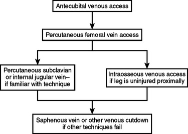
The ideal initial sites for vascular access in children are the peripheral veins in the upper extremities, especially the antecubital fossa. If access cannot be achieved in these vessels, central venous access may be employed (Figure 1). A percutaneous femoral venous catheter is the next best choice and the most commonly used route for emergency venous access in the child. This should be done without attempting a cut-down, preferably using the Seldinger technique. Surgeons familiar with subclavian catheterization in the child may utilize this route as the next choice.4 For surgeons comfortable with this technique in children, complications are rare. Intraosseous access is acceptable in injured children. Contraindications include proximal fractures and sites of infection nearby. The anteromedial surface of the proximal tibia is used, 2–4 cm distal to the tibial tuberosity. For insertion in the proximal tibia, the needle is directed inferiorly at a 45-degree angle from the perpendicular. If the insertion site is the distal tibia, the needle should be angled 45 degrees superiorly. In both instances, the goal is to angle away from the region of the growth plate and/or joint. There are specialized needles readily available to use with this technique, but if these are not available, a spinal needle with a trochar may be employed. Multiple entries into the medullary cavity should be avoided, as the leakage that occurs with multiple attempts may cause a compartment syndrome. For surgeons not familiar with peripheral, central, or intraosseous access in children, a cut-down of the saphenous vein may be employed,5 although intravenous (IV) access by cut-down is no easier or faster than any of the abovementioned methods.
Circulatory Management
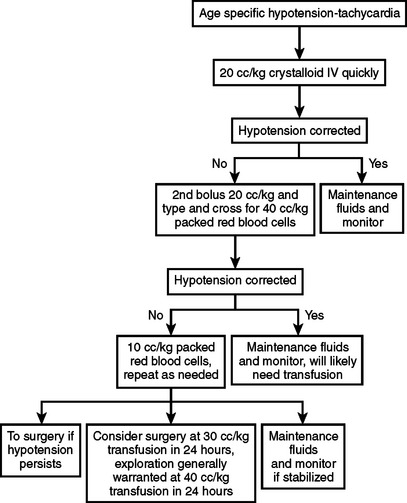
Age-specific hypotension is an indication for designating the necessity for major resuscitation in an injured child. In an analysis of the National Pediatric Trauma Registry, 38% of recorded deaths occurred in children whose systolic blood pressure was less than 90 mm Hg. This group represented 2.4% of the study population. To determine if a child has “age-specific hypotension” requires knowledge of normal blood pressures in children.6 New national guidelines for the ranges of normal childhood blood pressures based on age were published in 2004 (Table 1). A child with an injury that produces significant blood loss may present with a normal blood pressure. The otherwise healthy child can readily compensate for blood loss by mounting a significant tachycardia coupled with peripheral vasoconstriction. Therefore, a normal blood pressure in a child does not mean that circulating blood volume is at normal levels. A more accurate determination includes a blood pressure evaluation along with monitoring heart rate and assessing peripheral perfusion. Clinical signs of poor perfusion in conjunction with altered mentation are classic findings in pediatric hypovolemic shock. If these are present, then an immediate bolus of 20 cc/kg of isotonic crystalloid is in order. If a second bolus of this amount is needed, and there is little improvement, blood should be started immediately (Figure 2). Caution must be taken, as over-resuscitation may be as problematic as under-resuscitation, especially in the presence of a head injury. Enthusiastic administration of crystalloid solutions may exacerbate cerebral edema in certain circumstances. Overtreatment with crystalloid infusions may result in poor clot formation, worsening the compromised hemorrhagic state, and may have no impact on survival. Supranormal trauma resuscitation increases the likelihood of the abdominal compartment syndrome in adult trauma victims, and there are reports of the same problem in children.7 Typically, a bolus of 20 cc/kg of isotonic crystalloid in the presence of hypotension is the first treatment. If there is evidence of continuing instability, a second bolus of this amount may be given. If after two boluses of crystalloid the child does not have stable vital signs, blood should be started immediately. Type-specific packed red blood cells should be given, or O negative blood if necessary, in certain circumstances. Fresh frozen plasma and platelets should be considered early in the resuscitation period if large amounts of blood are needed for resuscitation. If there is a decreased level of consciousness without signs of hypovolemia, then modest fluid resuscitation is in order. Hypothermia is an extremely common occurrence in injured children. Hypothermia in the injured child may occur at any time of the year, even during the extremes of summer. The response to hypothermia includes catecholamine release and shivering, with an increase in oxygen consumption and metabolic acidosis. Hypothermia as well as acidosis contribute to a post-traumatic coagulopathy. Prevention and treatment of hypothermia require attention to this serious complication during the initial evaluation of the injured child. A warm room, warmed fluids, heated air-warming blankets, or externally warmed blankets should be utilized during the initial resuscitation. An aggressive approach to rewarming should begin in the emergency department and should be continued in the radiology suite during evaluation. There is some evidence to suggest that early, carefully controlled hypothermia in the severely head-injured child who has no other injuries may be beneficial, but this treatment option is still experimental.
Diagnostic Assessment
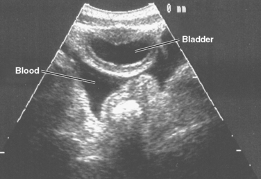
Several recent studies by adult and pediatric trauma surgeons have attempted to determine the role of focused acute sonography for trauma (FAST) in the evaluation of the injured child.8,9 The most common FAST evaluation examines the heart, right and left upper quadrants, and the pelvis for fluid (Figure 3). Some surgeons include an evaluation of the thorax for fluid in the pleural space and for pneumothorax.

Figure 3 A positive focused acute sonography for trauma (FAST) in a 4-year-old. Blood in the pelvis.
Stay updated, free articles. Join our Telegram channel

Full access? Get Clinical Tree


