Orthopedic Trauma, Fractures, and Dislocations
Samir Mehta
L. Scott Levin
I. Principles and Definitions of Orthopaedic Traumatic Injuries
Evaluation of the trauma patient with a potential fracture or dislocation should include a complete musculoskeletal evaluation, led by the mechanism of injury.
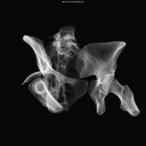
Dislocation is a complete loss of articular contact between two bones in a joint; this occurs with severe injury to ligamentous and capsular tissues. Define the direction of the dislocation with the distal piece described in relation to the proximal joint or bone (Fig. 31-1). Dislocations should be identified during the secondary survey and should be promptly reduced.
Findings on examination with joint dislocation include pain, loss of motion, shortening of the extremity, and associated neurovascular injuries (Table 31-1).
Reduction of major joint dislocation must occur as soon as possible after completion of a secondary survey. Reduction is performed with intravenous sedation, analgesia, and (in rare, major joints) chemical relaxation; use finesse rather than force. Occasionally, deep sedation or general anesthesia is needed for relocation.
Prereduction x-rays identify associated pathology. For example, a patient with a dislocated shoulder may have a non-displaced proximal humerus fracture, which may displace during reduction, leading to complications.
Subluxation refers to the partial loss of articular congruity; capsular and ligamentous structures remain to prevent complete dislocation. On a continuum, subluxation represents less injury than dislocation, although a complete dislocation that has partially reduced from the elastic recoil of the soft tissue can appear to be a subluxation.
A fracture is a structural break in bone continuity. Clinical signs include pain, displacement, shortening, swelling, and loss of function. Fractures are classified as either open (communicates with the external environment, Fig. 31-2) or closed. Describe the clinical deformity, soft tissues, and neurovascular structures, using this universal scheme.
The distal piece is described in relation to the proximal piece.
Fractures classifications include (Fig. 31-3):
Pattern. Transverse, oblique, spiral, other
Morphology. Simple (two parts) or comminuted (three or more parts)
Location. Proximal, middle, or distal; extraarticular or intraarticular
Radiographic parameters. Displacement, angulation, rotation, shortening, apposition
Assessing the degree of soft-tissue injury is important. Extensive soft-tissue injury increases the risk of the development of infection or compartment syndrome.
Pediatric fractures are classified according to their physical (growth plate) involvement (Salter–Harris classification).
Be familiar with the events surrounding the injury (mechanism of injury) and with the patient’s underlying medical conditions and current complaints. In the multiply injured patient, 15% to 20% of minor fractures (e.g., hand, foot, clavicle) are missed initially. The physical examination should:
Visually inspect for obvious soft-tissue abnormalities, including breaks in the skin, and deformities or asymmetry of the extremities.
Inspect the extremity throughout and turn the patient to the side (e.g., for spinal injuries, pelvic ring injuries extending to the anus).
Table 31-1 Neurovascular Injuries Associated with Fractures or Dislocations
Orthopedic injury
Neurovascular injury
Anterior shoulder dislocation
Axillary nerve injury, axillary artery injury
Humeral shaft fracture
Radial nerve injury
Supracondylar humeral fracture
Brachial artery, ulnar nerve injury
Distal radius fracture
Median nerve injury
Perilunate dislocation
Median nerve injury
Posterior hip dislocation
Sciatic nerve injury
Supracondylar femoral fracture/posterior
Popliteal artery injury/thrombosis
Knee dislocation/tibial plateau fracture/proximal fibular fracture
Peroneal nerve
Mangled extremity/tibial fracture
All neurovascular structures of the lower leg/compartment syndrome
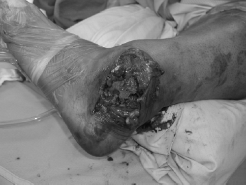
Figure 31-2. Open ankle fracture–dislocation with exposed bone and violation of the soft-tissue envelope.
Palpate bone and soft tissues to evaluate tenderness, crepitus, and firmness of compartments.
Test active and passive range of motion to detect bony injury, ligamentous injury, or weakness.
A neurovascular examination records quality of peripheral pulses, sensation (pinprick, light touch), and motor function.
Perform an additional tertiary orthopedic survey after the initial 24 to 48 hours or when the patient is more responsive to detect additional injuries.
Radiographic evaluation
Obtain at least two views at right (orthogonal) angles (generally anteroposterior and lateral views) of the involved extremity.
A traction x-ray of involved extremity may provide more information than a radiograph of displaced, subluxed, or dislocated joint. This is particularly helpful with the elbow (Fig. 31-4), distal femur, or the hip. In general, once the joint is reduced and out to length, computed tomography (CT) is essential to define periarticular fractures and to prepare for the operating room.
Imaging must include the joint above and the joint below the site of injury. For example, if the patient has a humeral shaft fracture, image the shoulder and elbow on the ipsilateral side.
Other views (e.g., stress views of the ankle or knee) may be necessary to assess joint stability.
Additional imaging, such as an MRI for a multi-ligamentous injury to the knee or arthrography for a shoulder dislocation, can be obtained later.
Emergent treatment of fractures or dislocations
Reduce the fracture or dislocation.
Splint/stabilize the extremity.
Irrigate open fractures with saline and cover with sterile saline-soaked gauze.
Administer parenteral antibiotics for open fractures or perioperatively in patients who require open reduction and internal fixation (ORIF).
Administer tetanus prophylaxis if >5 years since last dose for patients with open fractures or who require ORIF.
Definitive treatment of fractures or dislocations. The goal in treatment of musculoskeletal injury is to restore normal anatomy and function and relieve pain as quickly as possible. The early reduction and internal fixation in the multiply injured patient has reduced morbidity and mortality. The method of fixation should be adapted to the general condition (i.e., external fixation in unstable patients). Stabilization of the spine, pelvis, and long bone fractures (femur, tibia) allows early mobilization of patients.
Reduction can be accomplished by either closed (external realignment) or open (direct operative approach).
Immobilization of the extremity by splint, cast, traction, orthosis, external fixation, or internal fixation.
The most important measures in the care of open fractures are delivery of antibiotics, extensive and appropriate debridement followed by skeletal stabilization. Prevention of infection is of paramount importance. Development of osteomyelitis because of lack of antibiotic administration or inadequate debridement can be a devastating complication and will greatly increase the morbidity and potential for loss of function or limb. Antibiotics are not a substitute for effective debridement of necrotic and contaminated tissues.
Classification of open fractures (Gustilo and Anderson)
Type I. Low energy, <1 cm wound caused by protrusion of the bone through the skin or a low-velocity bullet
Type II. Moderate energy, >1 cm with flap or avulsion wound in the skin with minimal devitalized soft tissue and minimal contamination
Type III. High energy, extensive soft-tissue injury (usually >10 cm), “barnyard injury”
IIIa. Adequate soft-tissue coverage, no vascular injury necessitating repair
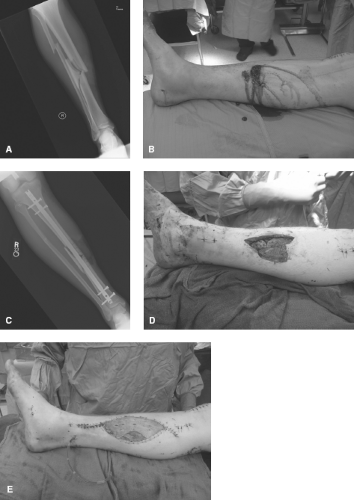
IIIb. Significant soft-tissue loss with exposed bone that requires tissue transfer for coverage (Fig. 31-5)
IIIc. Vascular injury requiring repair for limb preservation; amputation rates reported from 25% to 50%
Management after initial evaluation
Open fractures require urgent operative debridement and irrigation, with the orthopedic standard within 6 hours if the patient is physiologically stable.
Wounds require repeated irrigation and debridement in the operating room (OR) every 48 to 72 hours until definitive stabilization and soft-tissue coverage can be achieved (within 7 days of injury). Minimize multiple inspections of the wound outside the operating room except by the surgeon making critical management decisions. Continue antibiotics until 48 hours after definitive coverage.
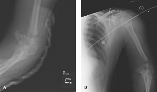
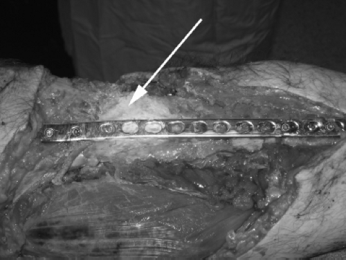
Antibiotic-impregnated bone cement “beads” and “blocks” may benefit delivery of antibiotics to spaces that do not receive the systemic antibiotics (Fig. 31-6). This local delivery has few systemic effects. The general mix dose is two vials of tobramycin (1.2 g each, 3.6 g total) per bag of cement and two vials of vancomycin (1 g each, 2 g total). The beads are strung on a stainless steel wire or heavy suture, allowed to harden, placed into the wound and covered with a liquid-sealed dressing (e.g., OpSite, Ioban). This “bead pouch” retains the fluid bathing the beads and is rich in antibiotic concentration.
Internal fixation of type I open fractures after adequate debridement and irrigation can be accomplished with infection rates of <10%. If soft-tissue loss is minimal and coverage is adequate, select type II and IIIa open fractures can be treated with intramedullary fixation. External fixation may be necessary to stabilize type III open fractures, where infection rates can range from 20% to 50%. A staged, planned redebridement may be necessary in open fractures with severe soft-tissue damage.
Wound closure may not be possible in all cases. Temporary closure with negative-pressure wound therapy may be required to provide temporary
coverage. At the end of the debridement, coverage of the bone is essential. However, negative-pressure should not be applied directly to exposed bone, tendon, nerve, or vessel. If adequate soft-tissue coverage cannot be achieved, an orthopedic or plastic surgeon skilled in managing extremity reconstruction should be consulted.
Gunshot injuries
Low-velocity gunshot injuries cause less soft-tissue destruction than high-velocity wounds. Because of the splintering effect of gunshot injuries, bone is often unstable and requires operative fixation. Debridement of the skin wounds and treatment of the bone as a closed injury is generally accepted. Prolonged antibiotic use is controversial; our practice is to administer 24 to 48 hours of intravenous antibiotics versus oral antibiotics for 5 to 7 days.
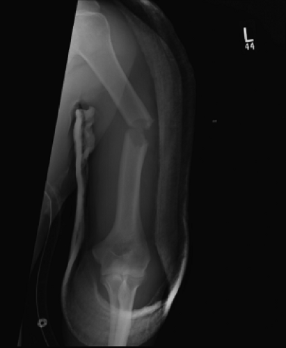
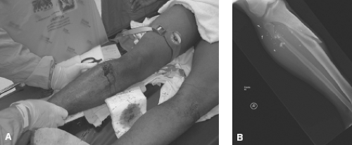
High-velocity gunshot wounds and close-range shotgun blasts cause significant soft-tissue injury, nerve and vascular injury, and should be treated as severe type III open fractures (Fig. 31-7). These wounds often require extensive reconstruction.
II. Identification and Management of Specific Orthopaedic Injuries
Pelvis
The pelvis is the supporting structure for the peritoneal contents and retroperitoneal structures. It connects the appendicular skeleton to the axial skeleton.
Because the pelvis lies in close proximity to vessels, the colon, and genitourinary structures, pelvic injuries can be associated with retroperitoneal bleeding and neurologic, bowel, and bladder injuries.
The sacrum and posterior ring are critical to the overall stability of the pelvic ring as the sacrum is the “keystone” to maintaining the biomechanics of ring congruity through force transmission.
Pelvic fractures may be defined as stable, rotationally unstable, or rotationally and vertically unstable.
All unstable injuries involve disruption of the posterior portion of the pelvic ring. Unstable pelvic fractures result from high-energy injury and are associated with 50% mortality in the multiple trauma patient. They require rapid assessment for stabilization and triage.
The anterior and the posterior pelvis should be inspected for open wounds. In males, the scrotal contents are palpated for testicular displacement and the penile meatus is examined for blood, which would suggest urethral injury. Assess rectal tone and possible laceration or prostate displacement. Women should undergo both bimanual and speculum examinations to rule out vaginal, urethral, and bladder injury. Vaginal or rectal laceration requires specific treatment.
Pelvic ring injury causing hypotension requires prompt diagnosis and treatment. Reducing the volume of the pelvis with a binding device (see later) is often effective in tamponading pelvic bleeding, which most commonly is from a venous source.
Posterior pelvic disruption can result in 3 to 4 L of blood loss and hemodynamic instability (Table 31-2). Concomitantly, aggressive intravenous resuscitation is necessary and may require blood product administration to achieve adequate hemodynamic stability. Patients who do not respond to resuscitative efforts should be continually re-evaluated to avoid a missed diagnosis for the underlying hypotension. If the working diagnosis remains hypotension secondary to pelvic ring disruption and hemorrhage, do angiography of the pelvic vasculature next after binding. In this scenario, a “blush” or active arterial bleeding source may be identified via angiogram and embolized at the time of the study. The most common source of arterial bleeding in the pelvis is injury to the superior gluteal artery.
Pelvic volume reduction (“binding”) can be done with a circumferential binder (either a bed sheet or a commercially available wrap [e.g., T-pod]) placed around
the pelvis and greater trochanters to reduce the intrapelvic volume (Fig. 31-8). It is imperative that the commercially available binders be assessed for soft-tissue pressure necrosis after every 24 hours. Percutaneous external fixation is a temporary measure before definitive ORIF. If pelvic stabilization is not possible or bleeding continues despite application of external fixation, angiography and embolization are therapeutic alternatives.
Table 31-2 Occult Blood Loss in Acute Fractures
Location of fracture
Blood loss (units)
Ankle
0.5–1.5
Elbow
0.5–1.5
Femur
1.0–2.0
Forearm
0.5–1.0
Hip
1.5–2.5
Humerus
1.0–2.0
Knee
1.0–1.5
Pelvis
1.5–4.5
Tibia
0.5–1.5
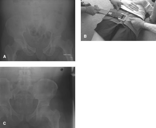
Figure 31-8. (A) AP pelvic radiograph of a 32-year-old patient who jumped from three-stories showing a disruption of the symphysis as well as widening of the sacroiliac joint. After application of a circumferential binder around the pelvis (B), the pelvis (and the pelvic volume) reduce dramatically as seen on the post-application AP pelvis radiograph (C).
Stay updated, free articles. Join our Telegram channel

Full access? Get Clinical Tree

 Get Clinical Tree app for offline access

