 Box 22.1 How to Test Visual Acuity
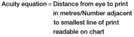
- Snellen letter chart is 6 m in front of patient (or equivalent distance using a mirror)
- Largest print on top line is usually 60
- Smallest print on bottom line is usually 5
- A patient with normal acuity can read down to line 6 and has ‘6/6’ vision
- With excellent acuity this may be ‘6/5’ vision
- A patient who cannot read the top line has vision ‘worse than 6/60’
- If the test is repeated at 3 m and the top line can now be read, vision is ‘3/60’
- The bigger the fraction, the better the vision
In the presence of blepharospasm (inability to open the eyes), further examination may require the application of local anaesthetic eyedrops, e.g. tetracaine (amethocaine) 1% or oxybuprocaine 0.4%.
- Evert the eyelids to allow examination of the fornices.
- Instil orange-coloured fluorescein drops to highlight any corneal damage – the injured epithelium appears green. A blue light makes a shallow pool of fluorescein much more easily visible.
- Examine the eye systematically from front to back using a slit-lamp and an ophthalmoscope. This may sometimes necessitate the administration of a short-acting mydriatic (a drug that dilates the pupil) such as tropicamide.
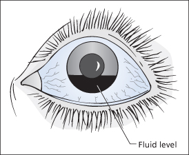
The anterior chamber may contain blood, which collects inferiorly in the erect patient to produce a fluid level. This haemorrhage is called a hyphaema (→ Figure 22.1). Damage to the lens or pupillary muscle will result in irregularities in the shape of the iris and disorganised or absent constriction to light. A laceration through the cornea will allow aqueous humour, muscle and even part of the lens to herniate anteriorly. Vitreous humour may be opacified by bleeding and the fundus thereby totally obscured.
Corneal Abrasions
Corneal abrasions may result from a scratch or other injury to the eye. Sometimes they follow prolonged rubbing of an irritating eye:
- The eye is red, injected, painful and watering.
- There is often pronounced blepharospasm (spasm of the eyelids).
- There may be a sensation of a foreign body.
TX
A foreign body must be excluded, especially a subtarsal one (→ below). The patient then requires the following:
- Dilatation of the pupil of the affected eye with a medium-duration mydriatic (e.g. cyclopentolate)
- Antibiotic ointment or viscous drops (e.g. fusidic acid) – three times a day for 3 days is sufficient and also easy to remember. The first dose should be instilled in the ED
- A firm eyepad overnight (→ Box 22.2)
- Instructions not to drive or operate machinery
- Either a review appointment or instructions to return if there is not a marked improvement within 24 h.
 Box 22.2 Padding an Eye
- A single pad will not keep an eye closed
- To apply a double pad, fold one pad in half and place it over the closed eyelids. Place a second, flat pad over the first and secure it in place with three pieces of tape
- The patient must be advised not to drive. Monocular vision impairs judgement of speed and distance. Patients who intend to drive (without a pad) must be instructed on how to apply the pads at home
- If both eyes require pads, leave this until the patient is at home
- All patients require spare pads and tape to take away
Most abrasions heal spontaneously within 2 days. Remember to check tetanus immunisation status.
Contact Lens Abrasions
Contact lens wearers may present with bilateral, large, shallow abrasions. There is often a history of prolonged use of hard lenses in a hot, dry, smoky atmosphere such as a nightclub.
TX
This is similar to the treatment for any other abrasion but is complicated by the fact that the lesions affect both eyes. Bilateral pads leave the patient without vision and can be deferred until safely home. Glasses should be used instead of contact lenses for at least a week. The patient should be advised to get a supply of artificial teardrops to lubricate the eyes whenever they are in similar circumstances to those in which the problem occurred.
Subtarsal Foreign Body
Foreign material may lodge in the conjunctival sac. There is an anatomical trap under the upper eyelid. The signs are those of corneal damage:
- The eye is red, injected and watering.
- There is pain and blepharospasm.
- There is a marked foreign body sensation.
- There is increased discomfort on blinking (but small children may keep their eyes tightly shut for relief).
- Linear scratches to the superior cornea are extremely suggestive of a subtarsal foreign body.
TX
Evert the eyelid, remove any foreign material, swab the tarsal conjunctiva and then treat as for a corneal abrasion. Local anaesthesia should be avoided if possible because the pain and foreign body sensation are dramatically relieved by the removal of the offending particles – a useful confirmation of the diagnosis.
Superficial Foreign Bodies Imbedded in the Eye
Fragments may fly into the eye while working (e.g. grinding) or may be blown in by the wind:
- The patient complains of a red, painful, watering eye.
- There is marked blepharospasm (it may be necessary to instil local anaesthetic drops before the eyelids can even be opened satisfactorily).
- The eye is injected and vision may be slightly blurred.
Always consider the possibility of deeper damage with higher-velocity injuries (→ below).
TX
Superficial fragments may be removed from the anaesthetised cornea using a cotton bud or a hypodermic needle. The resulting corneal ulcer should then be treated in the same way as a corneal abrasion.
Rust Rings:
metallic, ferrous, foreign bodies produce rust rings if left in the cornea for more than a few hours. If the rust is not removed completely, the patient should be reviewed at the eye clinic after 48 h when the softened area of damage will easily ‘shell out’.
‘Arc’ Eye
This occurs in a welder, skier or ultraviolet sunbather with inadequate eye protection. The condition is caused by corneal absorption of ultraviolet light. Problems usually start several hours after exposure:
- Both eyes may be affected.
- The eyes are painful, weeping and red.
- There is usually blepharospasm and a sensation of a foreign body.
- Fluorescein may reveal multiple tiny corneal erosions.
TX
Symptoms can be relieved by the instillation of local anaesthetic and dilatation of the pupil. The patient is then given topical antibiotics and an eye pad. The condition should settle within 48 h.
Chemical Splashes
Chemicals may cause inflammation of the conjunctiva or even a corneal burn. Patients present with an irritant or painful, watery, red eye. Alkalis may cause a penetrating eye injury.
TX
Irrigate the eye with copious amounts of saline. This is best achieved by emptying a 500-mL bag into the conjunctival sac through a standard giving set or by using a purpose-built irrigator. Ensure that both fornices are thoroughly washed and remove any lime or other fragments. Fluorescein drops will help reveal the extent of the damage. Antibiotic ointment should then be prescribed. Serious injuries must be referred to an ophthalmologist immediately.
Penetrating Injuries to the Eye
Deep, foreign bodies are difficult to identify, produce greater morbidity and yet, initially, may not be as painful as superficial lesions. A good history is essential. There is a high risk of intraocular damage in patients with eye pain who have worked with:
- a hammer and chisel
- glass
- machinery that emits high-speed fragments
- high-pressure water jets.
Fragments from grinding wheels rarely cause eye perforation.
The presence of hyphaema or prolapse of intraocular contents indicates severe injury, as does distortion of the pupil. Retained ferrous fragments will cause blindness because they are neurotoxic.
XR
Fluorescein-aided corneal examination is followed by radiology of the eye. Lateral views with the eye looking up and down (so that the foreign body appears to move) are particularly helpful in distinguishing foreign bodies from the normal bone pattern and from specks on the film.
TX
Immediate specialist referral is essential but acuity should be assessed first and an eye pad applied. Pressure on the eyeball and coughing and straining by the patient must be avoided for fear of dislodging intraocular structures.
Severe Blunt Trauma to the Eye
Damage to the globe of the eye most commonly results from sports injuries and assaults. Squash balls are particularly dangerous in this respect because they are small enough to enter the eye socket. Urgent ophthalmic referral is required in all cases. Pressure on the eyeball and coughing and straining by the patient must be avoided for fear of dislodging intraocular structures.
Hyphaema:
blood in the anterior chamber is caused by rupture of internal blood vessels (→ Figure 22.1). The patient is at risk of acute (secondary) glaucoma and requires bed rest and observation. A hyphaema may occasionally be accompanied by iridodialysis – rupture of the insertion of the iris.
Traumatic Mydriasis:
fixed dilatation of the pupil is a relatively common observation after blunt injury. The pupil may be distorted. Mydriasis is sometimes permanent. Traumatic miosis (constriction of the pupil) may also occur.
Dislocation of the Lens:
this is uncommon, although trauma is the most common cause. Cataract may also occur.
Posterior Segment Injuries:
the best sign of this type of trauma is a sudden reduction in visual acuity. Injuries include vitreous haemorrhage and retinal damage such as tears, haemorrhage and detachment. Choroidal rupture and even globe rupture are occasionally seen.
TX
The presence of any of the above conditions is an indication for an urgent ophthalmic opinion.
Subconjunctival Haemorrhage
Post-traumatic subconjunctival haemorrhage is common and usually trivial. Fluorescein may be used to reveal any conjunctival lacerations. Failure to visualise the posterior border of the haemorrhage may suggest orbital or retro-orbital trauma.
Spontaneous subconjunctival haemorrhage may follow coughing or straining. It is alarming to the patient but is usually of no consequence. Hypertension should be excluded.
TX
No specific treatment is required but the patient should be warned that the discoloration may take several weeks to resolve.
- Topical anaesthetics inhibit epithelial healing and protective ocular reflexes
- Topical steroids may exacerbate herpes infections or precipitate glaucoma
Assessment of the Red Eye
In the absence of a history of trauma, the cause of a red eye can be difficult to determine. Key observations are shown in Box 22.3. Minor trauma is the most common cause of a red eye in the ED.
 Box 22.3 Assessment of the Acute Red Eye: Key Observations to Help in Diagnosis
- Is the problem bilateral?
- Is there any associated visual loss?
- Is there a sensation of a foreign body?
- Is the eye uncomfortable and irritating or is it painful?
- Are the eyelids puffy or the conjunctiva oedematous?
- Is there any photophobia?
- Is the pupil irregular, dilated or unreactive?
Infective Conjunctivitis
This may be caused by several different agents:
- It is often bilateral.
- The eye is red, injected and uncomfortable with a gritty feeling.
Bacteria
cause the classic, sticky eye, which is worse on waking.
Viral conjunctivitis
results in itchy, watery eyes. There may be a concurrent upper respiratory tract infection (URTI). Injected follicular swelling in the lower fornix and a preauricular lymph node are characteristic findings
Adenovirus
causes a painful, puffy, watery red eye. The condition is slow to resolve and very infectious.
TX
As it may be difficult to differentiate between the types of infectious conjunctivitis, it is usual practice to treat them all with a topical antibiotic (e.g. chloramphenicol ointment or gentamicin eyedrops).
Allergic Conjunctivitis
This is caused by exposure to eyedrops, plants or other allergens, and is associated with atopy. Chloramphenicol, neomycin and atropine are the most common drug offenders:
- It is the most common cause of a puffy, red eye.
- There is lid swelling and conjunctival oedema (chemosis).
- It is irritating but not painful.
TX
Remove the offending substance. Eyedrops containing a vasoconstrictor and an antihistamine (e.g. Otrivine–Antistin drops) may be helpful.
Acute Conjunctival Oedema
Acute conjunctival oedema is sometimes seen without other signs of allergy, especially in children. The conjunctiva may balloon out of the lower fornix in a very alarming way.
TX
Local application of vasoconstrictor drops may be dramatically effective (→ before).
Episcleritis
This condition is idiopathic and self-limiting:
- It is uncomfortable and may be painful.
- A discrete area of conjunctiva is injected and swollen.
TX
Ophthalmological referral is required. NSAIDs can be helpful.
Orbital Cellulitis
This is most common in young children and arises from an infection of an adjacent sinus or eyelid. There may be a history of a URTI:
- The eye is red and the eyelids are puffy.
- Swelling may be painless at first but within hours there is pain, fever and distress.
TX
Admission and treatment with intravenous (IV) antibiotics is required. Referral may be made to an ENT (ear, nose and throat) surgeon, ophthalmologist or paediatrician, as suggested by the most likely origin of the infection and local policy.
Corneal Ulcers (Keratitis)
Dendritic ulcers:
these are caused by herpes simplex infection of the cornea:
- The eye is red and photophobic.
- There may be a foreign body sensation.
- There is a branching ulcer that stains with fluorescein.
Referral and therapy with aciclovir is required. Inappropriate steroid therapy can lead to corneal destruction and the need for a graft.
Bacterial corneal ulcers:
these may occur in patients with chronic corneal disease or corneal exposure as a result of lid disorders:
- The eye is red and painful.
- There is an intracorneal opacity.
The patient should be referred without antibiotic prescription.
Uveitis (Iritis)
This is an idiopathic inflammation of the anterior intraocular structures. It is often recurrent and in young adults may be associated with ankylosing spondylitis. There is:
- a red and painful eye with circumcorneal injection
- tenderness of the eye
- photophobia
- adhesions between the pupillary margin and the lens (synechiae), which may cause irregular dilatation of the pupil
- a haze over the cornea, especially the lower half.
TX
Local anaesthesia does not relieve symptoms; mydriasis may help. Urgent referral to an ophthalmologist is required.
Glaucoma
Acute angle-closure glaucoma is a disease of the over-40s and especially elderly people. It is associated with severe long-sightedness. Impaired outflow of aqueous humour from the anterior chamber of the eye causes raised intraocular pressure. Clinical features include the following:
- A red, painful eye, which is injected and tender and feels hard on palpation through the lid
- Visual loss, with the appearance of haloes around lights
- A semi-dilated, ovoid pupil, which does not react to direct or consensual light stimuli
- Corneal haze caused by oedema
- Malaise, nausea and vomiting.
TX
Immediate ophthalmic referral is essential. Discuss initial treatment that may include the following:
- Parenteral analgesics and antiemetics
- Pilocarpine eyedrops every 5 min for up to an hour
- Acetazolamide 500 mg orally or by slow IV injection
- Mannitol 20% (up to 500 mL) by slow IV infusion.
Recurrent Corneal Erosion
There is usually a history of a previous minor corneal injury. The patient describes a sharp discomfort on waking that settles after a few hours and then worsens over a few days. The signs are those of corneal damage.
TX
Referral and lubricating eye ointment at night for several months.
Marginal Keratitis
This is a recurrent condition. There is:
- a red, injected eye
- photophobia
- a sensation of a foreign body within the eye
- small, white patches within the cornea, close to the limbus, which do not stain well with fluorescein. These are sterile inflammatory infiltrates.
TX
Referral to the next eye clinic.
The Eyelids
Related posts:

Stay updated, free articles. Join our Telegram channel

Full access? Get Clinical Tree


