105 Neurologic Procedures
• Topical anesthetics (especially in children), liberal use of subcutaneous lidocaine, and small doses of anxiolytic agents can expedite performance of lumbar puncture in an anxious patient.
• To obtain an accurate opening pressure, patients should lie on their side with the head supported by a pillow and the legs straight. Cerebrospinal fluid should be allowed to equilibrate in the manometer before reading.
• A smaller-gauge needle with the bevel of the needle angled parallel to the dural fibers or use of a small-diameter blunt-tipped needle will decrease the frequency of post–dural puncture headache.
Lumbar Puncture
EPs perform LP to diagnose and treat a variety of neurologic complaints. Currently, this procedure is used mainly in the ED for assessment of headache or altered mental status. CSF analysis and opening pressure (OP) measurements obtained from the LP can make or exclude the diagnosis of meningitis, subarachnoid hemorrhage (SAH), pseudotumor cerebri, and other diseases with high morbidity. Although performance of LP is considered relatively safe, patients with a suspected mass lesion or ventricular obstruction may have a theoretic risk for herniation; in such cases neuroimaging may be warranted before LP.1
Procedure
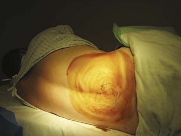
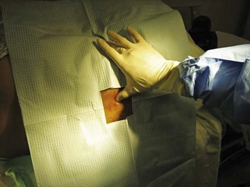
The iliac crest is located and the thumbs used to palpate and identify the spinous processes in the midline of the back. This is roughly the L4-L5 interspace and is an appropriate interspace to use. In adults, the L3-L4 interspace can also be used because both lie below the termination of the spinal cord within the spinal canal. An absorbent towel is placed under the patient, and the patient’s back is cleansed with the skin preparation solution (povidone-iodine or chlorhexidine) by making circular motions to scrub the back in gradually expanding circles, starting at the midline of the L4-L5 interspace and out to the iliac crests and lower thoracic spine; the skin should be allowed to dry between wipes (Figs. 105-1 and 105-2). The procedure may be facilitated by having the patient sit and lean over a bedside table while being stabilized by a helper, but this prevents measurement of OP. In anatomically challenging patients, ultrasonography and fluoroscopic guidance may aid in the identification of landmarks.
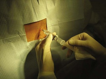
Once the patient and physician are in positions of comfort, the landmarks are reassessed and lidocaine is injected subcutaneously with a 27- or 25-gauge needle at the identified interspace to anesthetize the skin (Fig. 105.3). In children, topical application of anesthetic before injection may reduce the initial discomfort. After 1 to 2 minutes the deeper tissues are anesthetized by advancing a longer, 22-gauge needle slowly and injecting lidocaine. Achieving sufficient anesthesia is critical to success of the procedure.
Once anesthesia is achieved, the landmarks are checked yet again. A 22-gauge spinal needle is used to advance into the previously identified space (Fig. 105.4
Stay updated, free articles. Join our Telegram channel

Full access? Get Clinical Tree


