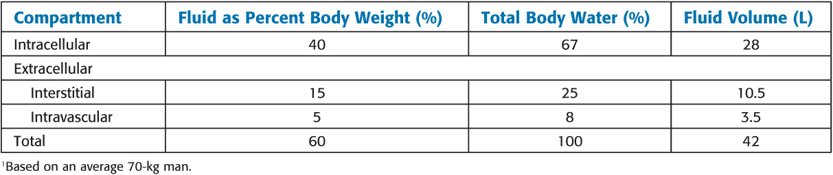
Intravascular fluid, commonly referred to as plasma, is restricted to the intravascular space by the vascular endothelium
![]() Most electrolytes (small ions) freely pass between plasma and the interstitium, resulting in nearly identical electrolyte composition
Most electrolytes (small ions) freely pass between plasma and the interstitium, resulting in nearly identical electrolyte composition
![]() Plasma proteins (mainly albumin) are the only osmotically active solutes in fluid not normally exchanged between plasma and interstitial fluid.
Plasma proteins (mainly albumin) are the only osmotically active solutes in fluid not normally exchanged between plasma and interstitial fluid.
Exchange Between Fluid Compartments
• Diffusion between interstitial fluid and ICF may take place by one of several mechanisms: (1) directly through the lipid bilayer of the cell membrane, (2) through protein channels within the membrane, or (3) by reversible binding to a carrier protein that can traverse the membrane (facilitated diffusion).
• Oxygen, CO2, water, and lipid-soluble molecules penetrate the cell membrane directly. Cations such as Na+, K+, and Ca2+ penetrate the membrane poorly because of the cell transmembrane voltage potential (which is positive to the outside) created by the Na+–K+ pump. Glucose and amino acids diffuse with the help of membrane-bound carrier proteins.
Disorders of Water Balance
Control of Plasma Osmolality
• Antidiuretic hormone
° When ECF osmolality increases, the hypothalamus releases antidiuretic hormone (ADH) from the posterior pituitary.
° ADH markedly increases water reabsorption in renal-collecting tubules
° Carotid baroreceptors and atrial stretch receptors can also stimulate ADH release after a 55% to 10% decrease in blood volume
• Thirst:
° Osmoreceptors in the lateral preoptic area of the hypothalamus are very sensitive to changes in extracellular osmolality. Activation of these neurons by increases in ECF osmolality induces thirst.
Hyperosmolality and Hypernatremia
• Hypernatremia and low total body sodium content
° Occurs when water loss is in relative excess to that of the sodium loss. Hypotonic losses can be renal (osmotic diuresis) or extrarenal (diarrhea or sweat).
° Urinary sodium concentration is generally greater than 20 mEq/L with renal losses and less than 10 mEq/L with extra-renal losses.
• Hypernatremia and normal total body sodium content
° Central diabetes insipidus
![]() Causes: Lesions involving hypothalamus and the pituitary, brain death, surgery
Causes: Lesions involving hypothalamus and the pituitary, brain death, surgery
![]() Diagnosis is suggested by a history of polydipsia and polyuria (often >6 L/d) and an absence of hyperglycemia. Diagnosis is confirmed by an increase in urinary osmolality after the administration of exogenous ADH.
Diagnosis is suggested by a history of polydipsia and polyuria (often >6 L/d) and an absence of hyperglycemia. Diagnosis is confirmed by an increase in urinary osmolality after the administration of exogenous ADH.
• Hypernatremia and normal total body sodium content
° Central diabetes insipidus (DI)
![]() Treatment: Aqueous vasopressin (5–10 U subcutaneously or intramuscularly every 4–6 hr) is the treatment of choice for acute central DI.
Treatment: Aqueous vasopressin (5–10 U subcutaneously or intramuscularly every 4–6 hr) is the treatment of choice for acute central DI.
![]() Desmopressin (DDAVP), a synthetic analogue of ADH with a 12- to 24-hour duration of action, is used both in the ambulatory and perioperative settings.
Desmopressin (DDAVP), a synthetic analogue of ADH with a 12- to 24-hour duration of action, is used both in the ambulatory and perioperative settings.
° Nephrogenic DI
![]() ADH secretion in nephrogenic DI is normal, but the kidneys fail to respond to ADH.
ADH secretion in nephrogenic DI is normal, but the kidneys fail to respond to ADH.
![]() Causes: Chronic renal disease, hypokalemia and hypercalcemia, sickle cell disease, and hyperproteinemias. Also can be secondary to the side effects of some drugs (amphotericin B, lithium, demeclocycline, ifosfamide, mannitol).
Causes: Chronic renal disease, hypokalemia and hypercalcemia, sickle cell disease, and hyperproteinemias. Also can be secondary to the side effects of some drugs (amphotericin B, lithium, demeclocycline, ifosfamide, mannitol).
![]() Diagnosis is confirmed by failure of the kidneys to produce a hypertonic urine after the administration of exogenous ADH.
Diagnosis is confirmed by failure of the kidneys to produce a hypertonic urine after the administration of exogenous ADH.
![]() Treatment is generally directed at the underlying illness and ensuring an adequate fluid intake.
Treatment is generally directed at the underlying illness and ensuring an adequate fluid intake.
• Hypernatremia and increased total body sodium content
° Most commonly results from the administration of large quantities of hypertonic saline solutions (3% NaCl or 7.5% NaHCO3)
° Primary hyperaldosteronism and Cushing syndrome may also have elevations in serum sodium concentration.
Treatment of Hypernatremia
• Rapid correction of hypernatremia can result in seizures, brain edema, permanent neurologic damage, and even death.
• In general, plasma sodium concentration should not be decreased faster than 0.5 mEq/L/hr.
Hyponatremia
Hypoosmolality and Hyponatremia
• Hypoosmolality is nearly always associated with hyponatremia ([Na+] <135 mEq/L).
• Pseudohyponatremia occurs when hyponatremia does not necessarily reflect hypoosmolality. Causes include hyperlipidemia, hyperproteinemia, glycine absorption during transurethral surgery, mannitol, and hyperglycemia.
• Hyponatremia invariably reflects water retention from either an absolute increase in total body water (TBW) or a loss of sodium in relative excess to loss of water.
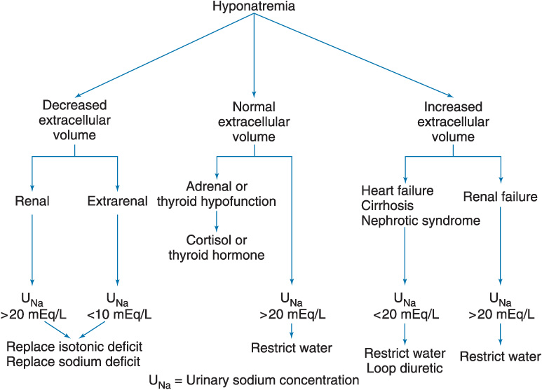
Hyponatremia and Low Total Body Sodium
• Loss of both sodium and water eventually lead to extracellular volume depletion. As the intravascular volume deficit reaches 5% to 10%, nonosmotic ADH secretion is activated.
• Causes include:
° Renal: thiazide diuretics, mineralocorticoid deficiency, salt-losing nephropathies, osmotic diuresis (glucose, mannitol), and renal tubular acidosis
° Extrarenal: Vomiting, diarrhea, integumentary loss (sweating, burns), and “third spacing”
Hyponatremia and Increased Total Body Sodium
• Edematous disorders are characterized by an increase in both total body sodium and TBW. When the increase in water exceeds that in sodium, hyponatremia occurs.
• Causes: Congestive heart failure, cirrhosis, renal failure, and nephrotic syndrome
Hyponatremia with Normal Total Body Sodium
• Hyponatremia in the absence of edema or hypovolemia may be seen with glucocorticoid insufficiency, hypothyroidism, drug therapy (chlorpropamide and cyclophosphamide), and the syndrome of inappropriate secretion of ADH (SIADH).
Clinical Manifestations of Hyponatremia
• Symptoms of hyponatremia are primarily neurologic and result from an increase in intracellular water.
• Patients with mild to moderate hyponatremia ([Na+] >125 mEq/L) are frequently asymptomatic.
• Early symptoms are typically nonspecific and may include anorexia, nausea, and weakness.
• Progressive cerebral edema results in lethargy, confusion, seizures, coma, and finally death.
Treatment of Hyponatremia
• Na+ deficit = TBW × (Desired [Na+] – Present [Na+])
• Very rapid correction of hyponatremia has been associated with demyelinating lesions in the pons (central pontine myelinolysis).
Anesthetic Considerations
• Plasma sodium concentration greater than 130 mEq/L is usually considered safe for patients undergoing general anesthesia.
• Plasma Na+ should be corrected to greater than 130 mEq/L for elective procedures even in the absence of neurologic symptoms
Disorders of Sodium Balance
Regulation of Sodium Balance and ECF volume
• Extracellular fluid volume and total body sodium content are ultimately controlled by appropriate adjustments in renal Na+ excretion.
Control Mechanisms
• Sensors of volume
° Baroreceptors at the carotid sinus and afferent renal arterioles (juxtaglomerular apparatus) indirectly function as sensors of intravascular volume.
° Whereas changes in blood pressure at the carotid sinus modulate sympathetic nervous system activity and nonosmotic ADH secretion, changes at the afferent renal arterioles modulate the renin–angiotensin–aldosterone system.
Related posts:
 Intravenous Anesthetics
Anesthesia for Patients with Cardiovascular Disease
Anesthesia for Genitourinary Surgery
Ambulatory, Non–Operating Room, and Office-Based Anesthesia
Intravenous Anesthetics
Anesthesia for Patients with Cardiovascular Disease
Anesthesia for Genitourinary Surgery
Ambulatory, Non–Operating Room, and Office-Based Anesthesia
 Spinal, Epidural, and Caudal Blocks
 Cholinesterase Inhibitors and Other Pharmacologic Antagonists to Neuromuscular Blocking Agents
Stay updated, free articles. Join our Telegram channel

Full access? Get Clinical Tree


