Anatomic Approach
Midline
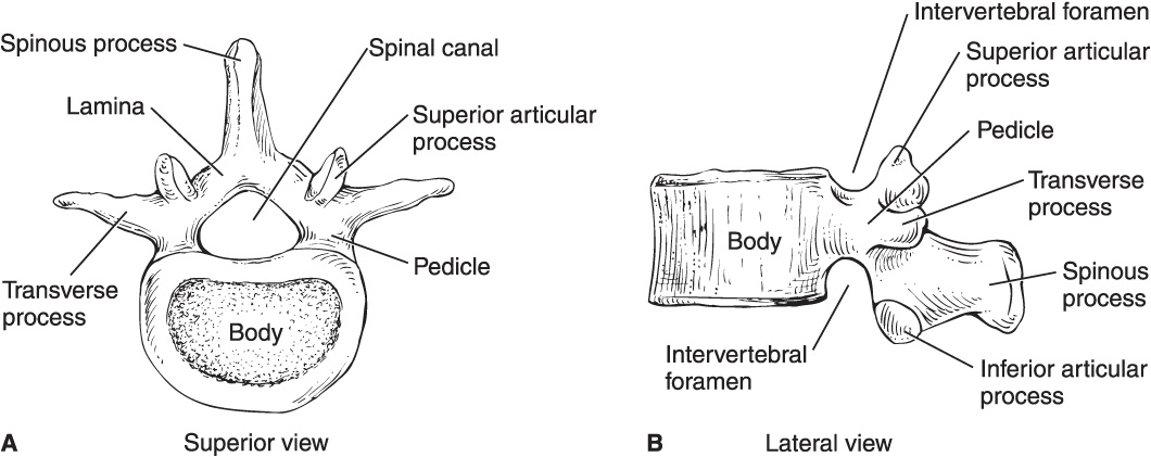
• Depression between spinous processes of the vertebra above and below the level to be used is palpated; this is the needle entry site.
• The needle is directed slightly cephalad.
• After passing through skin and subcutaneous tissue, the needle becomes engaged in supraspinous and intraspinous ligaments and eventually the ligamentum flavum.
• Epidural: Loss of resistance as the needle penetrates ligamentum flavum and enters the epidural space.
• Spinal: The needle is advanced through the epidural space and penetrates the dura-subarachnoid membranes.
Paramedian
• A paramedian approach is 2 cm lateral to the inferior aspect of the superior spinous process of the desired level.
• The needle is directed and advanced at a 10- to 25-degree angle toward the midline.
• Identification of the ligamentum flavum and entry into the epidural space is more subtle than in the midline approach.
Spinal Anesthesia (also referred to as Subarachnoid Block or Intrathecal Injection)
Spinal Needles
• Quincke: Cutting needle with end injection
• Whitacre: Pencil-point (round), side injection; lower risk of postdural puncture headache (PDPH)
• Sprotte: Side injection, long opening; more vigorous flow, but distal opening may not be in desired space, so full dose of medication may not be delivered
Technique for Spinal Anesthesia
• Median or paramedian approach
• Two pops: ligamentum flavum and dura-arachnoid membrane
• Successful dural puncture is verified by withdrawal of stylet and free flow of cerebrospinal fluid (CSF).
• Only preservative-free local anesthetics are used intrathecally.
• Vasoconstrictors (direct α-adrenergic agonist effect), epinephrine, and phenylephrine may be added to local anesthetic solutions; they decrease uptake or clearance from CSF and provide weak spinal analgesia.
• Opioids and clonidine (indirect, centrally acting α-adrenergic agonist) may be similarly added to enhance the quality and duration of spinal anesthesia.
• Hyperbaric bupivacaine and tetracaine are the most commonly used agents:
° Slow onset (5–10 min); prolonged duration (90–120 min)
• Lidocaine: Caution for transient neurologic symptoms (TNS) and cauda equina syndrome; may be safer if doses are limited to 60 mg and diluted to 2.5% or less with an opioid or CSF before injection
° Rapid onset (3–5 min); short duration (60–90 min)
Spinal Anesthesia
Factors Affecting Level of Block
• Higher dosage or site of injection generally leads to higher level of anesthesia.
• Baricity is relative to CSF specific gravity: Hyperbaric (denser) solutions spread according to gravitational pull (based on patient position), but the opposite is true for hypobaric (less dense) solutions.
° A head-up position causes a hyperbaric solution to settle caudad and a hypobaric solution to ascend cephalad.
° A supine position causes hyperbaric solutions to settle in the most dependent area of the spine (T4–T8 with normal thoracic kyphosis), thereby limiting spinal anesthesia to T4 and below.
• Local anesthetic solutions may be made hyperbaric by addition of glucose or hypobaric by addition of sterile water.
• Isobaric solutions remain at the level of injection; local anesthetic may be mixed with CSF (1:1) to make the solution isobaric.
• CSF volume inversely correlates with level of anesthesia; decreased CSF volume may be seen in elderly patients and in patients with severe kyphosis or kyphoscoliosis.
• CSF volume also decreased with increased intraabdominal pressure and engorgement of epidural veins: pregnancy, ascites, and large abdominal tumors.
• Decreased CSF volume means decreased dosage of spinal anesthetic to achieve the same level of blockade.
• It is unclear whether coughing or straining during injection affects spread of local anesthetic within the CSF.
Epidural Anesthesia
• May be performed at lumbar, thoracic, or cervical level
• Used for operative anesthesia, obstetric analgesia, postoperative pain control, and chronic pain management
• May use single-shot or catheter (with intermittent or continuous infusion)
• Epidural space includes fatty connective tissue, lymphatics, a venous plexus (Batson), and connective tissue septa
• Slower onset (10–20 min) compared with spinal
• Less dense than spinal; allows for differential blockade
• Segmental blockade: Confined relatively close to level of injection: well-defined band of anesthesia at certain nerve roots; nerve roots above and below are spared
• Lumbar epidurals are most common, least technically challenging, and overall safest
• Thoracic epidurals require greater angulation and carry an increased risk of potential spinal cord injury; paramedian approach often used
• Cervical epidurals primarily used for pain management
• 17 to 18 gauge
• 3 to 3.5 inches long
• Blunt bevel with curved (15–30 degree) tip; pushes away dura after passing through the ligamentum flavum
• Tuohy: Most common; Weiss = Tuohy with winged tip and introducer device set into hub
• Crawford: Straight; higher incidence of dural puncture, but easier to thread epidural catheter
Epidural catheters: Allow for continuous infusion or intermittent bolus techniques
• 19 to 20 gauge
• Advanced 2 to 6 cm into epidural space: shorter distance; more likely to dislodge, longer distance leads to a greater chance of unilateral block
• Single port at distal end or multiple side ports with a closed tip
• May be reinforced with spiral wire to resist kinking; associated with less intense paresthesia and may be associated with a lower incidence of inadvertent intravascular insertion
Techniques for Epidural Anesthesia
• Median or paramedian
• Loss of resistance to saline or air
• Hanging drop
Related posts:


Stay updated, free articles. Join our Telegram channel

Full access? Get Clinical Tree


