Fig. 76.1
Fundoscopic photograph showing intraretinal hemorrhage (blue arrow), tortuous blood vessel (black arrow), cotton wool spots (yellow arrow), and Roth spots (green arrow) (From Shirley and McNicholl [1]. Reprinted with permission from BMJ Publishing Group Ltd.)
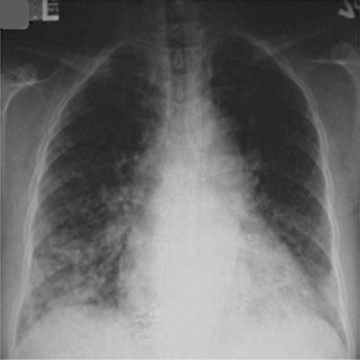
Fig. 76.2
Initial chest x-ray showing bilateral infiltrates in the lower lung zones (From Wu et al. [2]. Reprinted with permission from Elsevier Limited)
Question
What is the most likely diagnosis?
Answer
Hyperviscosity Syndrome due to Hyperleukocytosis
This patient exhibited the classic triad of mucosal bleeding, visual changes, and focal neurologic deficits seen in hyperviscosity syndromes. The elevated WBC with blasts on the differential suggested the possibility of an underlying hematologic malignancy as the etiology of the patient’s symptoms. In this case, emergent intubation was necessary given the patient’s impending hypoxic respiratory failure. Flow cytometry was performed and the patient was preliminarily diagnosed with acute myelogenous leukemia (AML). Oncology was consulted, who recommended emergent leukapheresis. After the leukapheresis, the patient markedly improved and was subsequently extubated. He later received two more sessions of leukapheresis resulting in complete resolution of his symptoms and a reduction in his WBC count to 33 thou/mcL. As seen in Fig. 76.3, the patient’s CXR also cleared completely after treatment. Bone marrow biopsy and flow cytometry confirmed the diagnosis of AML. The patient was started on chemotherapy and tolerated his medical treatment well and was later discharged with outpatient follow up of his AML.
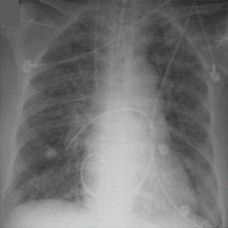
Fig. 76.3
Post chemotherapy CXR showing resolution of the patient’s lower lobe infiltrates (From Wu et al. [2]. Reprinted with permission from Elsevier Limited)
Principles of Management
Pathophysiology
Hyperviscosity syndrome (HVS) refers to the clinical constellation of symptoms exhibited due to an increase in serum viscosity. This increase in serum viscosity is the result of two major pathologic processes: most commonly paraproteinemias and less commonly excess cellular components (such as leukocytes and platelets). The resultant sluggish blood flow leads to a relative hypoperfusion and circulating proteins interfere with platelet aggregation causing prolonged bleeding time [3]. Classically, the patient will present with (among other symptoms) the triad of visual changes, mucosal bleeding, and focal neurological deficits [4]. Table 76.1 shows many of the end organ manifestations that can result from hyperviscosity syndromes.
Table 76.1
Clinical symptoms which can be seen in various hyperviscosity syndromes
Organ system | Hyperviscosity signs and symptoms | Diagnostic modalities |
|---|---|---|
Neurologic | Focal neurological deficits Headache Dizziness Altered Level of Consciousness Seizures Coma | CT Noncontrast Brain MRI Brain MRA Brain |
Pulmonary | Shortness of Breath Dyspnea Hypoxemia Wheezing | CXR CT Chest |
CVS | Chest Pain Palpitations Chest Tightness Acute MI | Cardiac Enzymes Echocardiogram |
Hematologic | Increased Bleeding Time Mucosal Bleeding Anemia Fatigue | CBC with diff PT/INR, PTT, fibrinogen, haptoglobin |
Ophthalmologic | Blurry Vision Retinal Vein Engorgement Progressive loss of sight Papilledema | Fundoscopic Exam |
In plasma, the main determinant of blood viscosity is protein concentration [5, 6]. Spherical proteins have a smaller effect on blood viscosity whereas larger proteins with high ratios of length to width (such as the pentameric IgM) can have an inordinate effect on viscosity [7]. This underlying mechanism explains the high incidence of HVS in lymphoplasmacytic lymphoma (formerly known as Waldenstroms Macrogloulinemia), a B cell lymphoma in which there are high levels of circulating IgM pentamers. The distribution of the gamma globulins also plays an important clinical role. IgM, predominantly an intravascular protein, exerts a greater effect on plasma viscosity compared to IgG and IgA (which are distributed more extravascularly) [5].
Leukostasis is the occlusion of blood vessels caused by hyperviscous blood due to an excessive number of circulating white blood cells. Hyperleukocytosis can be seen in both acute and chronic leukemias when the WBC is greater than 100,000 cells/mm3 and is more common when in their blast form [8]. Typically, it occurs in acute lymphoblastic leukemia (especially T cell variants), acute myeloid leukemia, and chronic myeloid leukemia during a blast crisis [8]. These immature blast cells (either myeloblasts or lymphoblasts) are larger and have a more rigid cellular membrane, making them more likely to cause leukostasis. Occlusion happens in small capillaries, and the excess WBCs release toxins that can damage the vascular endothelium resulting in local rupture and hemorrhage [8]. Leukostasis is a medical emergency when life-threatening symptoms develop and requires emergent cytoreduction. In our case, the patient exhibited hyperleukocytosis, as evidenced by his WBC of 145 thou/mcL and symptoms of leukostasis.
Diagnosis
The diagnosis of HVS requires a high degree of clinical suspicion supported by laboratory evidence showing an elevated protein level or hyperleukocytosis. Table 76.2 shows common cutoffs for which symptoms may be seen in both paraproteinemias as well as various blood disorders. It is important to emphasize that HVS remains primarily a clinical diagnosis that is additionally supported by laboratory data.
Table 76.2
A general guide for common cutoffs where the symptoms of hyperviscosity may present
Common cutoffs for hyperviscosity syndromes | ||
|---|---|---|
Paraproteinemia | IgM | >3 g/dL |
IgA | >10 g/dL | |
IgG | >15 g/dL | |
Excessive cellular components | AML | >300 thou/dL |
ALL | >600 thou/dL | |
CML, Blast Phase | >100 thou/dL | |
CML, Accelerated Phase | >100 thou/dL | |
CML, Chronic Phase (when pregnant) | >100 thou/dL | |
Essential Thrombocytosis | >1000 thou/dL | |
When one clinically suspects HVS due to paraproteinemia, the total protein level must be checked to see if it is elevated. If so, the next step is to perform a serum protein electrophoresis. HVS due to paraproteinemia is most commonly seen in Waldenstrom macroglobulinemia with an IgM level >3 g/dL [5]. In multiple myeloma with monoclonal IgG paraproteins, HVS does not appear until levels are >15 g/dL [5]. In IgA myeloma, the symptoms usually occur with plasma IgA levels >10 g/dL [5]. The intravascular distribution of IgM explains why symptoms of HVS occur at a much lower IgM protein level compared to both IgG and IgA paraproteinemias. In our case, the normal albumin-protein gap suggests that a paraproteinemia was not the underlying etiology of the patient’s symptoms and pointed more to hyperleukocytosis.
Related posts:


Stay updated, free articles. Join our Telegram channel

Full access? Get Clinical Tree


