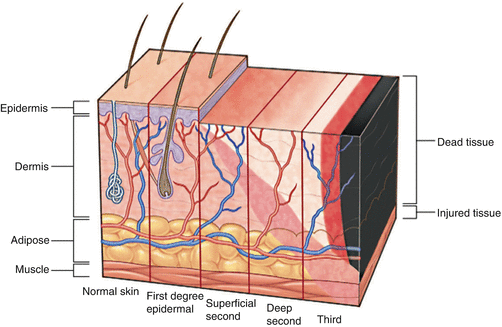
Fig. 83.1
A 35 year old male presents with partial and full thickness burns as depicted as a result of severe trauma and thermal injury. There is significant soft tissue damage, and eminent airway compromise as a result of the injury
Question
What is the most important first step in management?
Answer
In the setting of thermal injury, a complete primary survey must be performed in accordance with ACLS guidelines to assess the exact nature of injuries [1, 2]. On presentation, the airway must be secured, breathing must be assessed and large bore IV access must be placed to facilitate aggressive fluid resuscitation [3]. Though it is preferred to obtain IV access in non-burned tissues, sometimes this is necessary. Despite higher infectious risk, central access might still be required. Prior to presentation, it is of utmost importance to isolate the patient from the cause of the injury, using aggressive irrigation of the tissues if necessary in the setting of chemical injury.
This patient presents with facial burns, and it is critical to assess airway patency immediately on arrival. Bronchoscopy is the definitive form of airway assessment. Although it is also important to take note of physical exam findings that can indicate potential injury to the airway such as singed nasal hairs or soot in the airway, these exam findings are not always accurate, and early protection of the airway is critical to the immediate survival of patients presenting with thermal injuries [4]. If there is even minor concern for inhalation injury, immediate intubation is required. Chest X-ray can be performed at this time to ensure appropriate placement of the endotracheal tube and to serve as a baseline given that pneumonia is the most common inpatient complication and cause of death for patients with burns. Importantly, succinyl choline must be avoided on induction of anesthesia in patients with long-standing thermal injury given the high risk for hyperkalemia due to upregulation of acetylcholine receptors post-injury.
Once the airway has been secured and the patient is hemodynamically stable, the extent of burn injury must be quantified. Total body surface area (TBSA) is a critical factor in this process (Table 83.1). TBSA impacts survival. Patients with greater than 10 % TBSA burns are commonly admitted to an intensive care unit for monitoring. Importantly, only deep partial and full thickness burns are included in the determination of TBSA; superficial injuries are not included (Table 83.2). This patient has approximately 36 % TBSA burns (9 % anterior bilateral upper extremities, 9 % head/neck). There are three zones of tissue injury: 1. zone of coagulation, 2. zone of stasis, 3. zone of hyperemia. The zone of coagulation is the site most severely and irreversibly injured by the initial injury. The zone of stasis however, can be salvaged with appropriate resuscitation. The zone of hyperemia although damaged, can heal on its own without required additional interventions. Thus, immediate, aggressive resuscitation is critical to maximize tissue viability and salvage of the zone of stasis [1]. After the primary survey, the next step is to promptly initiate aggressive fluid resuscitation.
Table 83.1
Distribution of total body surface area
Anatomic surface | Percentage of total body surface area |
|---|---|
Head and neck | 9 % |
Anterior trunk | 18 % |
Posterior trunk | 18 % |
Upper extremities | 9 % each |
Lower extremities | 18 % each |
Genitalia | 1 % |
Table 83.2
Lund and Browder chart
Age (years) | 0–1 | 1–4 | 5–9 | 10–14 | 15 | Adult |
|---|---|---|---|---|---|---|
Head | 9.5 | 8.5 | 6.5 | 5.5 | 4.5 | 3.5 |
Anterior or Posterior Thigh | 2.75 | 3.25 | 4 | 4.5 | 4.5 | 4.75 |
Anterior or Posterior Leg | 2.5 | 2.5 | 2.75 | 3 | 3.25 | 3.5 |
Arm | 2 | 2 | 2 | 2 | 2 | 2 |
Forearm | 1.5 | 1.5 | 1.5 | 1.5 | 1.5 | 1.5 |
Hand | 1.5 | 1.5 | 1.5 | 1.5 | 1.5 | 1.5 |
Genitalia | 1 | 1 | 1 | 1 | 1 | 1 |
Gluteus | 5 | 5 | 5 | 5 | 5 | 5 |
Anterior or Posterior Thorax | 13 | 13 | 13 | 13 | 13 | 13 |
Neck | 1 | 1 | 1 | 1 | 1 | 1 |
Foot | 1.75 | 1.75 | 1.75 | 1.75 | 1.75 | 1.75 |
Principles of Management
Diagnosis
Several factors must be considered on initial evaluation of a burn. The type of burn (scald, electrical, chemical, or flame), depth of the burn (superficial partial, deep partial, full thickness), mechanism of injury, comorbidities, and social situation must be thoroughly investigated [5]. Superficial partial thickness burns penetrate through the superficial dermis, and are associated with severe pain given the lack of complete injury to the adnexal structures (Fig. 83.2). These burns commonly heal without surgical intervention. Deep partial thickness burns extend to the reticular dermis, and may not be associated with pain. Compared to the mildly hyperemic tissues of superficial partial thickness injuries, deep partial thickness burns are lobster red in color. Due to complete loss of adnexal structures, the tissues become dry, non-tender, and commonly require surgical intervention to ensure appropriate healing. Full thickness burns extend beyond the dermis into the subcutaneous tissues. Immediate surgical intervention is critical to optimal healing of these burns, and significantly minimizes the risk for burn wound cellulitis and sepsis [6].


