Classification
BMI range
Health risk
Overweight
25–30
Mild
Class I
30–35
Moderate
Class II
35–40
Severe
Class III
>40
Very severe
Respiratory Considerations
At baseline morbidly obese subjects may be mildly hypoxemic, with higher respiratory rates and lower tidal volumes. The compliance of the respiratory system is reduced resulting in increased work of breathing. Functional residual capacity (FRC) and expiratory reserve volume (ERV) decrease exponentially with increasing BMI , with the greatest rate of change in the overweight and mildly obese. In sitting subjects with a BMI of 30 kg/m2, FRC and ERV are only 75 % and 47 % of the values for person with a BMI of 20 kg/m2 [7]. In supine position, the effect of BMI on FRC is more pronounced and tidal volume may fall within closing capacity, promoting shunting.
The prevalence of sleep apnea in obese patients can be as high as 75 % [8]. Of those with sleep apnea and severe obesity up to 20 % may have the obesity hypoventilation syndrome (OHS) , which is characterized by awake hypercapnia, hypoxemia, and elevated HCO3 − [9]. It is important for anesthesiologists to recognize patients with OHS because it is associated with severe upper airway obstruction, restrictive lung disease, blunted central respiratory drive, and pulmonary hypertension.
High-volume injections of local anesthetics during neuraxial or brachial plexus blocks may further compromise the respiratory status. For example, an interscalene brachial plexus block may affect the phrenic nerve, leading to temporary paralysis of the ipsilateral hemidiaphragm. The use of low-volume ultrasound-guided interscalene block is associated with fewer respiratory complications with no change in postoperative analgesia compared with the standard-volume technique [10]. Spinal anesthesia in obese parturient scheduled for Caesarean section was associated with a BMI -dependent decrease of lung function, which persisted well into the recovery period, even longer than the actual presence of motor blockade [11]. In nonpregnant obese subjects, similar changes in lung function have been observed [12]. Intraoperative application of noninvasive positive pressure ventilation can improve respiratory function.
Cardiovascular Considerations
The increased tissue mass of the obese needs to be perfused leading to an increased total blood volume [13]. The increased total blood volume results in an increased cardiac output. Cardiac output increases from 4 L/min at a BMI of 20 kg/m2 to more than 6 L/min at BMIs greater than 40 kg/m2. Cardiac output affects the early pharmacokinetics, the front-end kinetics of drug distribution, and dilution in the first minutes after administration. An increased cardiac output decreases the fraction of drug distributed to the brain and increases the rate of redistribution, which may result in lower peak concentrations.
The most prevalent comorbidity in obese patients is hypertension. The increased cardiac output, the metabolic syndrome , diabetes, and physical inactivity all contribute to systolic and diastolic dysfunction even in otherwise healthy young obese subjects, which may eventually progress to left and/or right heart failure. The combination of super obesity (BMI >50 kg/m2) with hypertension and diabetes is associated with a twofold increased risk of death and adverse cardiac events in the perioperative phase [14]. There is evidence that epidural analgesia can reduce cardiovascular and pulmonary morbidity and mortality in high-risk obese patients undergoing major thoracic and abdominal surgery [15].
Pharmacology
Effect of Obesity
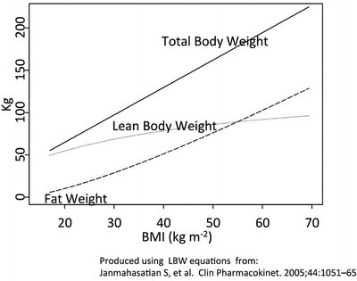
Until recently, obese subjects have been routinely excluded from clinical trials to obtain regulatory approval for investigational drugs. This has resulted in package insert dosage recommendations valid for normal weight patients but not for the obese. Obesity is not only associated with an increase in tissue mass but also changes in body composition and tissue perfusion. Fat mass and lean body mass both increase, but the increase is not proportional. The percentage of lean body mass as a percentage of total body weight decreases (Fig. 19.1). The different ratio of lean body weight to fat weight at different BMIs will have a significant impact on drug distribution. Fat perfusion is also altered at different BMIs. At low BMIs fat is relatively well perfused, at high BMIs fat is poorly perfused. Because of the different ratio of fat to lean body weight at different BMIs and changes in fat perfusion, the effect of obesity on drug distribution into the different tissues is poorly understood. The increased cardiac output of the obese decreases the fraction of drug distributed to the brain and increases the rate of redistribution, which may result in lower peak concentrations. In obese patients with normal cardiac function, cardiac output is highly correlated to lean body weight, more so than total body weight or other variables. Therefore, lean body weight and cardiac output are more appropriate dosing scalars than total body weight. Total body weight dosing will result in overdosing and side effects.


Fig. 19.1
Changes in body composition for a typical frame 167 cm tall female who increases her BMI . Lean body weight was calculated using the equations published by Janmahasatian, S., Duffull, S.B., Ash, S., Ward, L.C., Byrne, N.M. & Green, B. Quantification of lean bodyweight. Clin Pharmacokinet 44, 1051–65 (2005). Fat weigh was calculated by subtracting lean body weight from total body weight
Numerous pharmacokinetic studies have shown that clearance, the most relevant pharmacokinetic parameter for maintenance dosing is linearly related to lean body weight but not total body weight. This implies that lean body weight is the appropriate dosing scalar, not only for determining loading doses, but also for maintenance doses.
Sedation
Ideally administration of sedatives should be minimized or avoided. Respiratory depression caused by benzodiazepines and opioids is more pronounced in the obese, especially in those with obstructive sleep apnea . Upper airway collapsibility and decreased arousal response to airway occlusion make these patients particularly sensitive to drug-induced respiratory depression. Benzodiazepines decrease upper airway muscle activity with consequent obstruction and cause central apnea during the initial postadministration minutes. If very anxious, patients need premedication. Small doses of midazolam and opioids can be administered under continued monitoring. Recommend dosing scalars for several anesthetic agents used for sedation during block placement are summarized in Table 19.2.
Table 19.2
Recommended dosing scalars for IV sedative agents and opioids during regional anesthesia in obese patients
Sedative agents : | Dosing scalar | Comments |
|---|---|---|
Midazolam | LBW | Titrate very carefully, time to peak effect is 3 min |
Avoid in sleep apnea patients | ||
Synergistic respiratory depressant effect with opioids | ||
Cave airway obstruction | ||
Dexmedetomidine | LBW | |
Ketamine | LBW | Minimal respiratory depression |
Potent analgesic | ||
Propofol | LBW | For continuous infusion or maintenance dosing TBW |
Cave airway obstruction | ||
Opioids | ||
Fentanyl | LBW | Titrate to effect, time to peak effect is 3 min |
Alfentanil | LBW | |
Sufentanil | LBW | |
Remifentanil | LBW | TBW dosing may result in significant hypotension and/or bradycardia |
Local Anesthetics
Local anesthetics have a well-characterized side effect profile that includes the risk of CNS and cardiovascular toxicity and other adverse effects such as nerve injury and chondrolysis. There is no evidence that these side effects occur more often in the obese. In diabetic rats, an increase in nerve damage occurs after nerve block with traditional local anesthetics such as ropivacaine [16]. Some have suggested that patients with diabetes, a very common comorbidity associated with obesity, may increase the likelihood of nerve injury [17].
Studies specifically addressing the effect of obesity on local anesthetics are nonexisting, and consequently, the optimal dosing scalar for the administration of local anesthetics in obesity is not known. Because the clearance of many drugs in the obese is proportional to lean body weight, lean body weight dosing is probably more appropriate than total body weight dosing [18]. Continuous infusion regimens using total body weight may result in overdosing.
Lipid Rescue Therapy
The recommendation when a local anesthetic overdose is suspected is to administer 20 % lipid emulsion based on lean body weight: A dose of 1.5 mL/kg lean body weight of 20 % lipid emulsion delivered as a bolus over 1 min followed by a continuous infusion of 0.25 mL/kg/min for at least 10 min after return of spontaneous circulation. The bolus could be repeated once or the infusion doubled for continued hypotension, but the total dose, including both bolus and infusion, should not exceed 12 mL/kg [19].
Neuraxial Anesthesia
Epidural
Difficult Placement
Procedure times for epidural placement are longer in morbidly obese patients [20]. Anatomical landmarks may be difficult to identify and the depth from the skin to the epidural space is increased. Depth can increase from 3 cm at a BMI of 20 kg/m2 to more than 8 cm at BMIs greater than 40 kg/m2 [21]. Additionally, narrowed interspinous and interlaminar spaces as a result of degenerative spinal disease with ossification of the interspinous ligaments and hypertrophy of the facet joints may further complicate correct epidural placement.
Seventeen percent of morbidly obese parturients required a replacement epidural catheter due to inadequate pain control or failure to achieve adequate bilateral dermatomal sensory levels compared to 3 % in nonobese parturients [20]. Obese women are less able to identify the midline of their back accurately by touching with their finger compared with nonobese [22]. Half of the obese were accurate to within 5 mm in locating the middle of their back with their finger compared with 84 % nonobese women. Ultrasound can be used to identify the midline, the intervertebral space, and the distance from the skin to the epidural space. However, excess adipose tissue can impair identifying structures with ultrasound. Visualization of the spinous process and ligamentum flavum was estimated as “good” 70 % and 63 % of the time, respectively [23].
Accidental Dural Puncture
The incidence of complications with epidural anesthesia increases with increasing weight. In nonobese parturients, the incidence of accidental dural puncture, as a complication of epidural insertion for labor analgesia, has a reported incidence of 0.16–1.3 % [24]. In obese women, the incidence of accidental dural puncture has been reported to be as high as 4 % [25].
Postdural Puncture Headache
There is no evidence that obese women are less likely to develop a postdural puncture headache or that the characteristics of the headache and use of epidural blood patch are different [26].
Intravascular Puncture
Inadvertent epidural venous puncture occurs more frequently in the obese patient. One study reports a rate of 17 % vs. 3 % in nonobese [27].
Stay updated, free articles. Join our Telegram channel

Full access? Get Clinical Tree


