(1)
Chennai Breast Centre, Chennai, India
Invasive papillary carcinomas account for 1–2 % of all invasive breast carcinomas and tend to occur in postmenopausal women in their mid 60s. Papillary carcinomas can arise from benign papillomas and it is challenging to differentiate on a core biopsy. They are more prone to develop in women who have multiple peripheral papillomas than large centrally located intraduct papillomas.
Clinical Features
They often present as a well-circumscribed, soft masses in an elderly women. Some women may present with a bloody nipple discharge. Axillary lymphadenopathy is unusual.
Mammographic Features
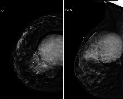
They are usually well-circumscribed, round or oval, lobulated hyperdense masses. Multiple masses may occur. Surrounding extensions and invasion may make the margins indistinct. There is associated papillary ductal carcinoma in situ which presents as microcalcifications within the solid component and the surrounding breast tissue (Fig. 36.1).


Fig. 36.1


Mammogram imaging showing a large dense circumscribed mass lesion
Stay updated, free articles. Join our Telegram channel

Full access? Get Clinical Tree


