(1)
Chennai Breast Centre, Chennai, India
Ultrasound correlation of mammographic abnormalities like discrete circumscribed masses, partially circumscribed masses, developing densities, asymmetric densities, and architectural distortion provides a more specific diagnosis of mammographic abnormality. Ultrasound can reliably distinguish cysts from solid lesions. Therefore, ultrasound can downgrade BI-RADS 3 masses on mammography to BI-RADS 2 on ultrasound, thereby obviating the need for a 6-month follow-up. Correlating mammographic abnormality with ultrasound also offers easier and quicker access to biopsy or localizes the lesion for surgical excision. Ultrasound is useful to assess the extent of disease and assess the axillary lymph nodes when mammographic appearances are strongly suggestive of a malignancy.
During ultrasound examination, it is essential to ensure that the abnormality seen on ultrasound is the same as the mammography abnormality. The size, shape, location, and surrounding tissue echogenicity should correlate with that of mammographic abnormality.
The size of lesion seen on the mammogram usually correlates well with ultrasound. Sometimes, the lesions may then appear larger on mammography than on ultrasound. This is because the glandular density and mass lesion or any abnormality appears as white density on mammogram, and when there is no intervening fat in between the normal glandular tissue and the abnormality, there is summation effect making it appear larger. However on ultrasound, the abnormal tissues have a different echogenicity (hypo- or hyperechogenicity) from the surrounding normal tissue.
The shape of a lesion on mammogram and ultrasound can be different because of the differences in compression and rotation maneuvers involved in performing the imaging. Compressibility of lesion also influences the shape of the lesion. Hence, a vertically ovoid lesion on a mammogram can be transversely ovoid on ultrasound. Compressible lesion like a cyst will appear spherical on mammogram but elliptical on ultrasound (Fig. 13.1).
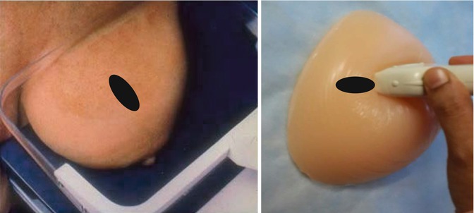
Fig. 13.1


Mammography pulls the breast away from the chest wall, whereas ultrasound compresses the breast against the chest wall. In a compressible lesion, the orientation of the lesion varies accordingly
Related posts:


Stay updated, free articles. Join our Telegram channel
Full access? Get Clinical Tree
