(1)
Chennai Breast Centre, Chennai, India
Breast Ultrasound: Equipment Settings and Examination Technique
Equipment settings have a critical influence in breast ultrasound. The examination must be performed with a B-mode imager. A linear array transducer in the frequency range of 7.5–13 MHz is recommended. As the operating frequency increases, the resolution improves but the imaging depth decreases. The 7.5 MHz provides a good trade-off between the resolution and the depth (Fig. 11.1).
Fig. 11.1
Linear array transducers
Modern ultrasound units can be preset to any of the several basic settings and require only minor adjustments. A high dynamic range (50–70 dB) with medium contrast and a high frame rate and line density is recommended.
The TGC slope including the near and far field should be tailored to each patient by adjusting the side switches so that the fat is medium gray from the subcutaneous tissue to the chest wall. Incorrect gain may make a solid lesion appear cystic and vice versa (Figs. 11.2 and 11.3a, b).
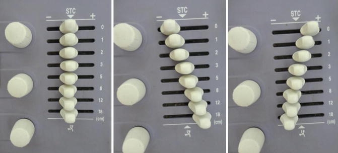
Fig. 11.2
TGS setting
Fig. 11.3
(a) Image on the right showing correct gain settings. The fat is medium gray from the subcutaneous tissue to the chest wall. The cyst appears anechoic with posterior enhancement. (b) The image on the left shows incorrect gain settings with artifactual echoes within the simple cyst
A penetration depth of at least 4 cm with selectable focal regions is required. The field of vision should include the entire breast from the skin to the ribs. For a more detailed examination, a smaller field may be selected (Fig. 11.4a, b). A transducer that provides 2–3 focal zones is useful, but having more than three focal zones reduces the frame rate. When the frame rate is reduced, the image generation lags behind the scanning and is no longer real time (Fig. 11.5).

Fig. 11.4
(a, b) Image on the left shows inadequate depth settings. The retromammary region of the breast is not completely included in the image. The image on the right shows proper adequate depth settings with the retromammary portion of the breast included while scanning
Fig. 11.5
(a, b) The image on the left shows focal point incorrectly placed. (b) the image on the right shows focal point placed correctly in the region of interest. the lesion is visualized better
Split screen imaging is a very useful tool in breast ultrasound. It is useful in documenting lesions in both transverse and longitudinal or radial and antiradial scans. It is also useful to compare mirror images on both sides of the breast to evaluate an asymmetry or a palpable thickening. Split screen images can also stitch up images to increase the field of view twice the width of the long axis of the probe in situations where the lesion is larger than the standard transducer (Fig. 11.6a, b).
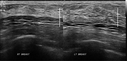
Fig. 11.6
Split screen images showing asymmetric glandular tissue on the right side. RT Right, LT Left
A larger field of view can also be obtained by other methods. A 5-MHz curved linear transducer can be used. The length of the transducer can be increased to 50 mm instead of the standard 38-mm transducer. The 50-mm transducer will also speed up the scanning time in larger breasts. A virtual convex image or trapezoidal image can also be used to increase the field of view. The center of the image maintains the lateral resolution, but the lateral resolution is reduced in the periphery by phasing and divergence of the beam. This trapezoidal image can show the entire margin of larger lesion’s and measure its largest dimension (Fig. 11.7).
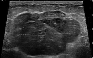
Fig. 11.7
A virtual convex image or trapezoid image can also be used to increase the field of view
For very large lesions in the breast, it is possible to image the entire lesion by panoramic image (Fig. 11.8) or by using a low-frequency ultrasound probe (Fig. 11.9).
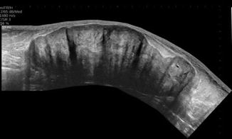
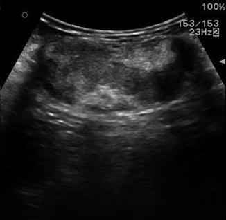
Fig. 11.8
A panaromic view can also be used to increase the field of view
Fig. 11.9
A low frequency curvilinear probe can also be used to visualized a large lesion
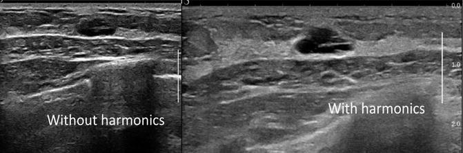
Modern high-frequency machines are equipped with digitally encoded harmonics, which reduce the speckle artifact. Calcifications can be seen better with the reduction of speckle artifact. Harmonics reduces the artefactual echoes within the lesion, and therefore there is a greater clarity in differentiating complex from simple cysts (Fig. 11.10). It also helps to define the thin echogenic pseudocapsule that surrounds most benign solid and complex cystic lesions within the breast, because it improves the axial resolution. Reduced penetration and reduced frame rate particularly when used in conjunction with Doppler are the limitations with harmonics. In a woman with large and dense breast, fundamental imaging without harmonics will be required to ensure that the deeper tissues are visualized. It may also be necessary to turn off the harmonics when Doppler examination is being carried out to maintain the frame rate.