Inflammation


Inflammation


LOCAL PROCESS

Inflammation presumably evolved as a process to achieve two principle benefits—wound healing and defense against microbiologic invasion. When insults are relatively small (i.e., elective groin hernia wound, facial acne), then the physiology and cellular functions of these two processes can be considered local, somewhat distinct, with little effect on the rest of the body (little systemic effect). But when insults are large (i.e., bilateral femur fractures with bilateral pulmonary contusions, perforated sigmoid diverticulitis), then the similarities between wound healing and host-defense mechanism become more apparent and associated with more evidence of systemic alterations.

First, here are descriptions of the more local physiology and cellular functions of wound healing and host defense, followed by descriptions of how wounds and microbiologic invasion result in systemic inflammation.


Wound Healing

When a wound is in the skin and subcutaneous tissue (the most common wound studied), the epidermal barrier is broken and keratinocytes release pre-stored interleukin-1 (IL-1). The subsequent response is bleeding and coagulation. Platelet activation results in the release of important chemoattractants [i.e., epidermal growth factor (EGF), platelet-derived growth factor (PDGF), transforming growth factor-beta (TGF-β)].

Damaged blood vessels initially constrict, but this is soon followed by vasodilation and increased capillary permeability secondary to the action of agents such as prostaglandin E2, prostacyclin, histamine, serotonin, and kinin. This vascular response results in the accumulation of protein-rich edema fluid (exudate). Leukocyte cells adhere to the damaged and leaky vessels.

Attracted by chemoattractants such as PDGF and IL-1, polymorphonuclear cells (PMNs) are the first leukocytes to migrate to the inflammatory site (within minutes if the circulation is good). PMNs serve to phagocytize dead tissue and foreign objects. Removal of bacteria may be assisted by opsonins and preformed antibodies. PMNs produce proteases and intracellular oxygen radicals that are critical for beneficial PMN activity. Besides proteases and oxygen radicals, PMNs can release IL-1. IL-8 is a potent PMN attractant produced by many cell types after incubation with IL-1 and tumor necrosis factor (TNF). The PMNs last only for hours.

Lymphocytes are the next cell to migrate into the wound. The role of the lymphocyte is less well understood, but depletion of T cells results in impaired wound healing.

Subsequently (within hours), tissue macrophages and circulating monocytes are attracted by substances such as PDGF, TGF-β, and IL-1, migrate into the injured area, and last for days to weeks. Wounds can heal without PMNs but not without macrophages that regulate most of the continuing stages of inflammation and wound healing through mediators such as IL-1, PDGF, TGF-β, TGF-α, and fibroblast growth factor (FGF).

Fibroblasts migration and angiogenesis begin next. Fibroblasts are influenced by IL-1, PDGF, TGF-β, and TGF-α; angiogenesis is influenced by TGF-β, TGF-α, and EGF. The combined process of fibroblast proliferation and capillary budding produces a wound that is granular in appearance (granulation tissue), very vascular, and quite friable. Certain fibroblasts—myofibroblasts—have smooth muscle contractile elements that contract and diminish the area of a wound. In general, wound contraction continues until the lining cells from each edge of a wound meet (epithelialization for the skin). Therefore, slight contraction will follow wounds that are closed primarily, that is, with the lining edges apposed. Much contraction may occur in secondary healing, especially when wound edges remain widely separated for days to weeks.


Fibroblasts make collagen, usually accelerating at five days after tissue damage. Before this, fibrin is the principle wound substance besides sutures that holds a wound together. Collagen synthesis is also influenced by IL-1, PDGF, TGF-β, and EGF. Since the macrophage is an important source of these factors, mechanisms that increase (glucan administration) and decrease (the combined effect of hypoxia and endotoxin) macrophage function may result in increased or decreased wound strength, respectively.

A summary of cellular activity in wound healing is provided in Tables 4.1 and 4.2 (1, 2).


Local Host Defense

Local host-defense mechanisms have been principally studied as the response to microbiologic challenge. Both an innate immune capacity (defenses that lack immunologic memory) and an acquired capacity (defenses with immunologic memory) are components of local host defense (Tables 4.3 and 4.4).

The first mechanism of local innate host defense is surface barrier function, best understood as the effect of the intact skin. Penetration through a barrier results in further stimulation of the innate response.

The polymorphonuclear leukocyte (PMN, neutrophil) is the earliest cell component that responds to pathogen penetration. The steps related to PMN infiltration and action are listed in Table 4.3. PMNs serve to phagocytose organisms and/or debris, kill microorganisms, and release enzymes and reactive oxygen species to enhance control of damaging materials. Complement coating of organisms enhances phagocytosis (3, 4, 5, 6, 7).

PMN activation also results in the release of cytokines (IL-1, IL-6, TNF-α, IL-8, IL-12), toxic oxygen radicals, peroxides, nitric oxide (NO), and lipid mediators of inflammation such as prostaglandins and leukotrienes, as well as platelet activating factor (PAF) (5).


Table 4.1 Normal Wound Healing/Normal Inflammation


Events


Cells responsible


  • Coagulation


  • Early inflammation


  • Later inflammation


  • Collagen and mucopolysaccharide


  • Capillary budding


  • Wound contraction


  • Collagen remodeling


  • Platelets


  • PMN (first few hours)


  • Monocytes (days) macrophages


  • Fibroblasts (maximum deposition 7-10 days)


  • Endothelial cells (maximum 7-10 days)


  • Myofibroblasts


  • Fewer fibroblasts and capillaries


Table 4.2 Functions of Wound Healing Cells


Cells


Function


• Keratinocytes


• Release IL-1


• Platelets


• Coagulation, release EGF, PDGF, TGF-β, VEGF


• PMN


• Phagocytosis, especially microbes, release IL-1, IL-8


• Macrophage


• Phagocytosis, stimulate fibroblast migration and growth, stimulate endothelial cell migration and growth, release FGF, PDGF, IL-1, IL-6, TGF-β, TGF-α


• Fibroblast


• Collagen deposition, TGF-β wound contraction, TGF-β, PDGF wound remodeling, FGF, TGF-β


• Endothelial cells


• Capillary budding, VEGF


Abbreviations: IL-1, interleukin 1; EGF, epidermal growth factor; PDGF, platelet-derived growth factor; TGF-β, transforming growth factor beta; VEGF, vascular endothelial growth factor; IL-8, interleukin 8; FGF, fibroblast growth factor; IL-6, interleukin 6; TGF-α, transforming growth factor alpha.


Macrophage infiltration follows PMN activation and accomplishes many of the same microbe suppression activities. In addition, macrophages are responsive to pathogen-associated molecular structures (PAMP) that are shared by entire classes of pathogens, such as lipopoly-saccharides on Gram-negative bacteria. Macrophage recognition of a PAMP triggers activation before proliferation at the site (8). The macrophage also serves as an antigen-presenting cell for stimulation of the acquired immune system. However, the dendritic cell, also responsive to PAMP, is a more potent antigen-presenting cell that migrates to local lymph nodes to engage T cells (7).

The principal PAMP receptors are called toll-like receptors, first described in drosophila, and now considered a primary mechanism for activation of nuclear factor-kappa B (NF-κB), an important step in the production of cytokines and co-stimulatory molecules (8, 9, 10).

Natural killer cells can recognize “non-self” cells by absence of the major histocompatibility complex (MHC) class I. Since MHC class I is present on nucleated cells, most are not subject to threat. However, infection can alter the expression of MHC class I on infected cells and result in killer cell destruction (7, 11).


Table 4.3 Components of Local Host Defense: Innate Response


  1. Surface barrier


    1. Epithelial cells


      1. Skin


      2. Mucosa


    2. Constituents


      1. Mucus


      2. Normal flora


  2. PMNs


    1. Margination


    2. Rolling


    3. Adhesion to endothelial cells


    4. Transmigration


    5. Phagocytosis


    6. Release of toxic molecules


  3. Macrophages


    1. Phagocytosis


    2. Stimulation of angiogenesis


    3. Stimulation of contraction


  4. Dendritic cells—presentation of antigens


  5. Natural killer cells—augment removal of “non-self”


  6. Complement activation


    1. Opsonization


    2. Stimulation of inflammatory cells


  7. Systemic alterations in the “local” response


    1. Fever


    2. Acute phase reactants


Table 4.4 Components of Local Host Defense: The Acquired Response


  1. Antigen presentation


    1. Dendritic cells


    2. Macrophages


    3. B cells


  2. T-cell activation


  3. T-cell differentiation


    1. TH1—cellular immunity


    2. TH2—humoral immunity


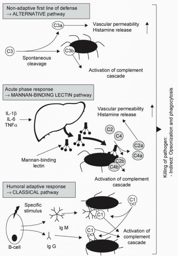
Complement is another component of the innate immune system that can be activated via three pathways (Figure 4.1). The classical pathway is activated by antibodies [immunoglobulin (IgG, IgM] binding to antigen and is part of acquired immunity. The mannan-binding lectin (MBL) pathway is activated by binding of MBL, an acute-phase reactant secreted by the liver, to bacteria or virus surface components. In the alternative pathway, the complement component C3 becomes “spontaneously activated” to C3b that then binds to the surface of a pathogen and promotes opsonization. Overall, activation of complement results in opsonization, recruitment of inflammatory cells, and direct killing of pathogens (5).

Small amounts of tissue injury and infection can presumably result in principally autocrine and/or paracrine effects that do not result in any cellular activity distant from the site of the stimulus. However, sufficient tissue injury and infection initiates an endocrine effect such
that mediators of inflammation cause systemic alterations that can be adaptive or maladaptive, usually depending on the magnitude of insult and, sometimes, the magnitude of the individual’s response (see section on “Severe Systemic Inflammation”).


Figure 4.1 Complement activation at different time points following bacterial invasion. First is the alternative pathway that is triggered by binding of C3 to the surface of a pathogen. Second, the acute-phase response results in a mannan-binding lectin that can bind to bacteria and activate complement. Finally, the classic pathway can be triggered by specific antibody that has accrued against an organism. Source: From Ref. 5.

Adaptive systemic effects that appear to enhance host defenses are fever (secondary to IL-1, IL-6, and TNF-α) and liver synthesis of acute phase reactants [i.e., C-reactive protein (CRP) and MBL], which are enhanced by these same cytokines (5).

As with wound healing, the local response to pathogen penetration can result in complete resolution of tissue injury with little scar formation. However, incomplete resolution can result in ongoing inflammation, granulation tissue formation, and fibrosis, collectively called an abscess. An abscess may or may not be associated with systemic alterations, again dependent on the magnitude of the threat as well as that of individual response.

Acquired immunity depends on the presentation of foreign antigens to T cells. Dendritic cells, macrophages, and B cells serve to present antigen to unstimulated T cells. Antigens that gain access to the circulation can be captured by antigen-presenting cells in the spleen and stimulate T cell activation at that site. The naïve T cells can then differentiate into Th1 cells that augment cell-mediated immunity or Th2 cells that augment humoral immunity. This differentiation is linked to a complex interaction of inflammatory mediators. Full activation of acquired immunity takes several days (5).

In addition to these inflammatory cell activities, a local site of infection is characterized by arteriolar vasodilation, increased blood flow, increased capillary permeability, exudation of plasma, and pain. This results in the complaint of pain (dolor), and the findings of erythema (rubor), edema (tumor), and warmth (calor).

Stimulants to Host-Defense Responses


Alarmins

The recognition that the inflammatory response to wounded tissue is similar to cell processes during infection has highlighted that host defense is not strictly limited to recognition of “self” from “non-self.” Perhaps the most striking example of variability in recognition of “self” is the difference in inflammatory response to programmed cell death (apoptosis) and cell disruption by injury and/or ischemia (necrosis/oncosis). Apoptosis results in little inflammatory response, but necrosis incites a response as vigorous as wounding or infection. Unprogrammed cell death (necrosis) releases molecules (collectively termed “alarmins”) that are not released during apoptosis, can be augmented by inflammatory cell activity, and can result in activation of receptor-sensitive cells that enhance acquired immunity. Molecules that have been catalogued as alarmins include the following: High Mobility Group Box 1 (HMGB1), a nuclear protein; heat-shock proteins (HSP); uric acid. Alarmins and PAMP have been combined under the term damage-associated molecular patterns (DAMP) to enhance the consideration that tissue injury and infection can cause similar host-defense responses (9).


Ischemia/Reperfusion

Oxidative stress is defined as a state of excess reactive oxygen intermediates (ROIs) compared with the endogenous antioxidants of the host. Oxidative stress can result from an excess of ROI production or a deficit in antioxidants. The production of ROI is one mechanism whereby phagocytes (PMNs and macrophages) kill invading organisms, an obvious benefit. However, while intracellular ROI can provide potent host-defense activity, extracellular ROI can cause injury to host tissues and also augment the local and systemic inflammatory response (12).

Phagocytic cell activation is not the only mechanism for excess ROI production. Ischemia results in depletion of cellular ATP and accumulation of xanthine and hypoxanthine as ATP is metabolized. Two intracellular enzymes [xanthine dehydrogenase (XD) and xanthine oxidase (XO)] can metabolize these breakdown products, with XD using NAD+ as an electron acceptor, but XO using molecular oxygen. Cell energy deficits augment calcium influx into the cell that increases conversion of XD to XO. When oxygen is re-introduced (reperfusion), the action of XO results in the accumulation of superoxide anion and hydrogen peroxide, both toxic moieties to host cells (12, 13, 14).


Table 4.5 Oxidative Stress Molecules


  1. Superoxide (O2−•)


  2. Hydrogen peroxide (H2O2)


  3. Hydroxiy radical (HO)


  4. Hypochlorous acid (HOCL)


  5. Peroxynitrate (ONOO)


While PMNs are required for the full expression of ischemia/reperfusion injury, endothelial cells appear to be particularly capable of ischemia/reperfusion ROI production, even in the absence of neutrophils. The ubiquitous presence of endothelial cells provides equally ubiquitous potential for nearby cell damage via oxidative stress mechanisms.

Molecules that participate in oxidative stress states are listed in Table 4.5. Additional mechanisms for ROI production include nitric oxide metabolism and lipid peroxidation as a feature of cell membrane damage. Interestingly, lipid peroxidation is also a principle mechanism of cell injury from ROI, illustrating the potential for repetitive ROI injury. Cell death from oxidative stress appears to be linked to excessive mitochondrial calcium accumulation (13).


Ischemia Alone (Hypoperfusion Begets Inflammation)

Insufficient oxygen delivery can cause activation of inflammatory cells and other innate host-defense mechanisms before reperfusion. Mountain sickness is associated with increased capillary permeability (lungs, brain) and elevated blood concentrations of IL-6 and CRP. Hypoxia has been shown to increase the activity of innate immune cells, and PMN activation has been documented during hemorrhage and during cardiopulmonary resuscitation. Finally, rapid restoration of the circulation (i.e., rapid improvement in delivery of oxygen to cells, before reperfusion mechanisms are activated) has been shown to diminish markers of inflammation (15, 16, 17, 18).


SYSTEMIC PROCESS

While it is common for mild-to-moderate inflammatory conditions to result in some systemic manifestations (e.g., fever, tachycardia, leukocytosis, increased blood levels of CRP), more severe inflammatory conditions can cause life-threatening functional disturbances in organs distant from the principle inflammatory site, meeting the definition of severe systemic inflammation. In 1992, the concept of systemic inflammatory response syndrome (SIRS) was offered in keeping with the recognition that tissue injury with and without infection was capable of producing systemic alterations. The diagnostic criteria for SIRS are listed in Table 4.6 (19). While this listing included variables typically associated with severe, organ function-threatening systemic inflammation (i.e., hypothermia and leukopenia), the cataloguing of SIRS reflected severity only as related to infection (sepsis, severe sepsis, septic shock) and did not provide a more generic ranking of the severity of systemic inflammation per se. Since then, the magnitude of systemic inflammation-associated organ malfunction (MODS scores, SOFA scores) has been used to monitor severity and can, therefore, be used to identify severe systemic inflammation, regardless of etiology.

The magnitude of the systemic response to an inflammatory stimulus appears to be linked to two principle pathophysiologic mechanisms that cause cell function abnormalities: circulation deficits (too little oxygen delivery to cells); an excess of inflammatory toxins (cytokines, oxidative stress molecules, etc.). While each of these processes can be a primary manifestation of systemic inflammation, it is characteristic for both to be acting simultaneously to threaten cell function and/or viability.


Circulation Deficits (Inflammation Begets Hypoperfusion)

Mechanisms of inflammation-induced hypoperfusion are listed in Table 4.7. Vasodilation and increased vascular capacitance have been linked to activation of ATP-sensitive potassium channels
(from decreased intracellular ATP), the efflux of potassium from the cell, hyperpolarization of the cell membrane, and inhibition of calcium entry into the cell. Since augmented cytosolic calcium concentrations cause smooth muscle contraction, the inhibition of calcium entry limits vasoconstriction. Inflammatory stimuli increase the activity of inducible, calcium-independent, nitric oxide synthetase in endothelial and smooth muscle cells, resulting in vasodilation that is resistant to catecholamines and angiotensin II. In addition, a vasopressin deficiency (i.e., concentrations insufficient to produce arteriolar constriction) has been documented in prolonged hypotension following both hemorrhagic and inflammation-induced hypoperfusion (20, 21, 22, 23).


Table 4.6 Systemic Inflammatory Response Syndrome


  1. Body temperature >38°C or <36°C


  2. Heart rate greater than 90 BPM


  3. Tachypnea, manifested by a respiratory rate >20 breaths per minute, or hyperventilation, as indicated by a PaCO2 of <32 mm Hg


  4. An alteration in white blood cell count, such as a count >12,000/mm3, a count <4,000/mm3, or the presence of more than 10% immature neutrophils


Table 4.7 Mechanisms of Inflammation Induced Hypoperfusion


  1. Increased capacitance (vasodilation), decreased MCFP, decreased venous return


    1. Hyperpolarization of vascular smooth muscle membrane


    2. Increased nitric oxide concentrations


    3. Decreased vasopressin concentrations


  2. Decreased vascular volume (exudation of plasma, decreased ECF—“third space” losses


    1. Exudation at the site of tissue injury


    2. Exudation at sites distant from site of injury


    3. Fluid accumulation in the lumen of the GIT


    4. Cell membrane deficits—migration of ECF into the intracellular space


  3. Impairment of the microcirculation


    1. Regional vasodilation/vasoconstriction


    2. Plugging—leukocytes/platelets


  4. Myocardial depression


    1. Ischemia—microcirculation


    2. Myocardial depressant factors


Abbreviations: MCFP, mean circulatory flow pressure; ECF, extracellular fluid; GIT, gastrointestinal tract.


Venous return is also threatened by a decrease in plasma volume from the migration of both plasma and interstitial fluid into sites collectively called “the third space.” The exudation of plasma at the site of injury and/or infection is an obvious process of plasma volume depletion. But the other components of extracellular fluid sequestration (exudation at other sites, fluid accumulation in the gastrointestinal lumen, and enhanced intracellular water migration) are not so intuitive. Increased systemic capillary permeability is presumably secondary to the endocrine effects of inflammatory mediators on endothelial cells throughout the body, an effect most regularly witnessed during alterations in lung function, yet possible to identify in the glomerular capillary (24, 25, 26, 27). Intestinal alterations are common with acute illness. The balance between secretion and absorption can be altered, resulting in further depletion of plasma volume (28). When inflammation is sufficiently severe, cell energetics and cell membrane function can be impaired, disturbing membrane pump function that maintains the normal concentrations of intracellular versus extracellular ions. Sodium pump deficits will result in increased intracellular sodium concentrations, causing migration of interstitial water into the intracellular space (29, 30).

Systemic inflammation can result in marked alterations in the microcirculation, including constriction of larger arterioles, dilation of smaller arterioles, alterations in capillary flow, and plugging by leukocytes and platelets. These deficits may variously affect specific tissues. The intestinal tract appears to be particularly sensitive. Recently, the measurement of sublingual microvascular flow in human sepsis has demonstrated that the potential for marked reduction
in regional oxygen delivery is present even when global (i.e., cardiac output-based oxygen delivery) is excellent (31, 32, 33).

Myocardial depression is a common feature of severe systemic inflammation, even when the overall circulatory state is hyperdynamic (34). Systolic malfunction such as a global reduction in ejection fraction and/or regional wall motion abnormalities as well as diastolic dysfunction have been documented with echocardiography (35). Both cardiac troponin and NH2 terminal pro-brain natriuretic peptide (NT-proBNP) elevations parallel the severity of depression as well as the prognosis (36, 37, 38). The mechanism(s) responsible for these alterations are imperfectly characterized, but include microcirculation ischemia and myocardial depressant factors that encompass a host of molecular mediators (34, 35, 39). Importantly, measurement of an elevated cardiac troponin or NT-proBNP does not necessarily “rule in” other cardiac diagnoses associated with these biomarkers, especially when severe systemic inflammation is present.


Excess Inflammatory Toxins

As mentioned in the Shock chapter, deficits in cell metabolism have been linked to toxic mechanisms that can inhibit cell function separately from oxygen metabolic pathways. The term cytopathic hypoxia has been applied to these metabolic alterations that mimic the changes from oxygen deficits (40, 41). Severe systemic inflammation appears to be the principal clinical cause of cytopathic hypoxia. Several studies in humans have documented that decreased oxygen consumption and/or an inability to increase oxygen consumption following resuscitation are associated with high mortality (42, 43, 44). In essence, these patients exhibit continuing evidence of the Ebb Phase of shock despite efforts that improve total body oxygen delivery.

The concept of toxic cellular injury is supported by epidemiologic data showing higher concentrations of inflammatory biomarkers in the most severely ill patients (45, 46, 47, 48). In addition, human studies have periodically demonstrated good tissue oxygen concentrations despite evidence of organ malfunction (49, 50).

The molecular etiologies of cytopathic hypoxic have been associated with diminished delivery of pyruvate into the mitochondrial tricarboxylic acid cycle, inhibition of key mitochondrial enzymes, and activation of the enzyme poly(ADP-ribose) polymerase-1 (PARP-1) (40).

In addition to the potential for inflammatory toxins to cause metabolic alterations, the products associated with oxidative stress can cause direct cellular injury as described above (12). PMN activation is the primary mechanism for oxidative tissue injury and augmented PMN activation with tissue sequestration has been linked to multiorgan malfunction in both experimental and human investigations (51).


METABOLIC AND HORMONAL RESPONSE TO INFLAMMATION

The metabolic and hormonal response in severe tissue injury and systemic inflammation is linked to the Ebb and Flow phases of shock (Table 4.8) (52). As noted in the Shock chapter, persistence of the Ebb phase is usually lethal. Achievement of the Flow phase is associated with increased temperature, resting energy expenditure (REE), oxygen utilization, and glucose and fat oxidation. Amino acids are also used as substrate, and there is an increase in nitrogen loss. At three to four days following injury, the Flow phase can be at a maximum and then diminish gradually in association with resolution of ileus, spontaneous diuresis of “third space,” and normothermia (53). When inflammation continues, most commonly secondary to new or unresolved infection, persistence of the Flow phase can be associated with progressive organ failure and death, sometimes despite the eradication of the principal inflammatory focus (53).


Carbohydrate

Glucose is commonly elevated in both the Ebb and Flow phases. In the Ebb phase, the glycolytic and glucogenic endocrine response is associated with a poor insulin secretion (54). During the Flow phase, the combination of increased epinephrine, cortisol, glucagon, lactate, and release of glucogenic amino acids from muscle (especially alanine) results in elevated glucose, often despite increased blood insulin concentrations. With progressing systemic inflammation, glucose intolerance increases, with gluconeogenesis accelerated by mass action of lactate and
alanine. Glucose uptake is augmented, particularly at major inflammatory foci, such as a large wound. Therefore, elevated glucose production may provide an important energy substrate for inflammatory cells (52, 53, 55, 56).


Table 4.8 Summary of Metabolic Changes During Trauma and Severe Systemic Inflammation in Humans


Severe Inflammation


Substance


Trauma


EBB


Flow


Adrenaline


INC


N


INC


Noradrenaline


INC


N


INC


Cortisol


INC


N


INC


Insulin


DEC


INC


VAR


Glucose


INC


VAR


VAR


Fatty acids


DEC


VAR


DEC


Albumin


VAR


DEC


DEC


Acute reactants


VAR


INC


INC


Urine nitrogen


VAR


INC


INC


Ree


VAR


INC


INC


Glucose oxidation


DEC


INC


DEC


Fat oxidation


INC


INC


INC


Abbreviations: N, normal; INC, increased; DEC, decreased; VAR, variable.

Only gold members can continue reading. Log In or Register to continue

Stay updated, free articles. Join our Telegram channel

Jul 5, 2016 | Posted by in CRITICAL CARE | Comments Off on Inflammation

Full access? Get Clinical Tree

Get Clinical Tree app for offline access