Chapter 41
Increased Intracranial Pressure
An acute rise in intracranial pressure (ICP) in patients in the intensive care unit (ICU) is a life-threatening cranial compartment syndrome that requires emergent treatment. In adults, the cranial contents are bounded by a rigid skull, which is noncompliant and unable to accommodate additional volume. Intracranial hypertension becomes deleterious when it reduces cerebral blood flow or when it results in mechanical compression of brain tissue. Clinically, intracranial hypertension is often inferred from neurologic examination, from changes in vital signs, or from findings on neuroimaging studies. ICP may be measured directly during lumbar puncture or with a variety of ICP monitors. Standardized indications for ICP monitoring have been established by the Brain Trauma Foundation for patients with traumatic brain injury (Chapter 99), but they are lacking in other disease states. Initial therapy for acute intracranial hypertension is often empiric, but its cause should be sought immediately.
Physiology
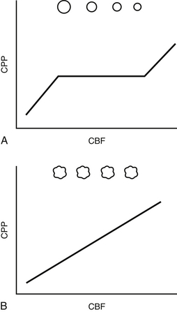
The goal of resuscitation in the neurocritical care patient is to provide sufficient delivery of fuel, typically oxygen and glucose, to meet cerebral cellular metabolic demand. Fuel delivery is largely determined by cerebral blood flow (CBF), which is difficult to measure directly at the bedside. Cerebral perfusion pressure (CPP) is therefore commonly used as a surrogate for CBF. CPP is calculated as the difference between mean arterial pressure (MAP) and ICP. The relationship between CBF and CPP may be approximated using Poiseuille’s law, which describes flow through a rigid tube. Modeled this way, CBF is directly proportional to CPP and to the vessel radius raised to the fourth power, and it is inversely proportional to blood viscosity. Under normal circumstances, cerebral autoregulation maintains constant CBF across a wide range of CPP through modulation of vascular diameter. In many disease states (e.g., traumatic brain injury), however, vasoplegia and impaired autoregulation develop, disrupting this relationship (Figure 41.1). When this occurs, CBF becomes linearly related to CPP (CBF becomes “pressure passive”), and it becomes crucial to closely monitor and tightly control CPP. This necessitates measurement and control of ICP.
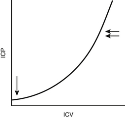
The Monro-Kellie doctrine defines intracranial pressure (ICP) as the sum of the pressures exerted by the intracranial contents, namely brain tissue, blood, and cerebrospinal fluid (CSF). It is intuitive that each of these components exerts pressure because of its volume. However, the relationship between intracranial volume and intracranial pressure is nonlinear. The intracranial compliance (change in volume per unit change in pressure) curve is logarithmic (Figure 41.2). The intracranial compartment may absorb from 100 to 150 mL of additional volume without a significant change in ICP because of a variety of compensatory mechanisms such as collapse of the low-pressure veins and egress of CSF into the lumbar subarachnoid space. Once these compensatory mechanisms are exhausted, however, additional volume results in a rapid rise in ICP. Normal ICP is roughly 6 to 12 mm Hg, and, by convention, intracranial hypertension is defined as ICP > 20 mm Hg.
The average volume of CSF in the adult is ~140 mL, of which ~20% is in the ventricular system, 60% is in the intracranial subarachnoid space, and 20% is in the spinal subarachnoid space. CSF is an ultrafiltrate of plasma and is largely produced by the choroid plexus, focal invaginations of the pia and its vessels into the lumen of the ventricles. CSF production is normally balanced by CSF reabsorption. When CSF exits the ventricles, it flows into the subarachnoid space and is absorbed into venous blood via arachnoid villi, small projections of arachnoid within the cerebral dural sinuses.
Pathophysiology and Differential Diagnosis
The causes of intracranial hypertension may be divided mechanistically into disorders of CSF dynamics, hydrocephalus, cerebral edema, and mass lesions. Box 41.1 provides a limited differential diagnosis for intracranial hypertension, categorized by cause.
< div class='tao-gold-member'>
Stay updated, free articles. Join our Telegram channel

Full access? Get Clinical Tree


